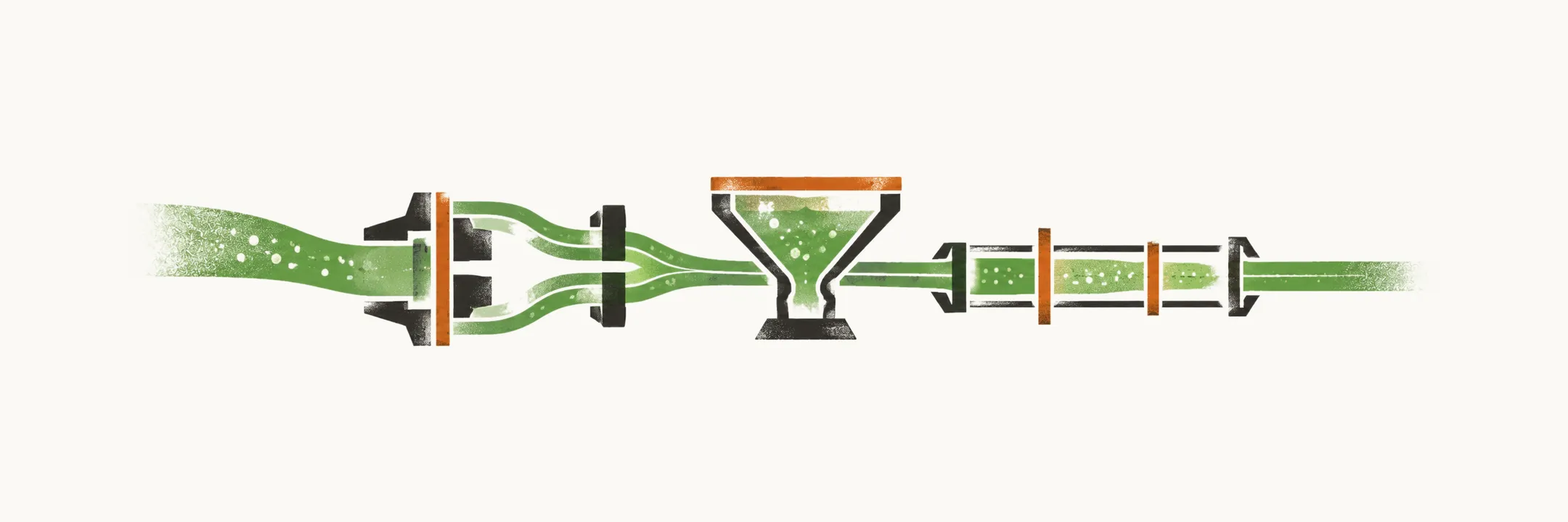
Hyponatremia is a plasma sodium concentration below the normal range. The work of diagnosis is to decide which of the three groups from Causes of Hyponatremia the patient belongs to: intravascular volume depletion; hypotonic fluid intake that exceeds the kidney’s water excretion; or the syndrome of inappropriate antidiuresis (SIAD). In SIAD, vasopressin (AVP, also called ADH) keeps acting when it should be suppressed, as ADH and the Regulation of Plasma Tonicity explains.
So the picture is a sorting. The bedside places the patient in a group, and the laboratory tests then check that placement, one question each. Management follows from two things: how severe the presentation is, and what the cause is.
Why do the history and examination come first?
History and physical examination are the most important step in the diagnosis and differential diagnosis of hyponatremia. They decide which of the three causal groups the patient belongs to before any laboratory result returns. The history looks for the routes by which water is gained or lost, and for the drugs and diseases that drive each group. What does it ask?
What does the history ask?
The history asks about each group in turn.
- Fluid loss, for example vomiting or diarrhea. Fluid loss and diuretics point toward hypovolemia, because low volume keeps ADH on, so water is held and sodium keeps falling.
- The causes of SIAD. Drugs, CNS, pulmonary or malignant disease point toward SIAD.
- Symptoms of endocrine dysfunction that suggest hypoadrenalism or hypopituitarism.
- Medication and drug use.
- Psychological conditions such as psychogenic polydipsia. Excessive thirst points toward water intake that exceeds excretion.
What does the examination check?
The examination asks the same question at the bedside, and it fixes the volume state. The body judges volume by how full the arteries are, so the signs are read with that in mind. It looks for:
- signs of extracellular volume depletion;
- orthostatic or persistent hypotension;
- signs of peripheral oedema or ascites;
- signs of heart failure, cirrhosis or renal failure.
The last two groups of signs matter because a body can look full of fluid and still act as if its arteries were short.
What do the laboratory tests answer?
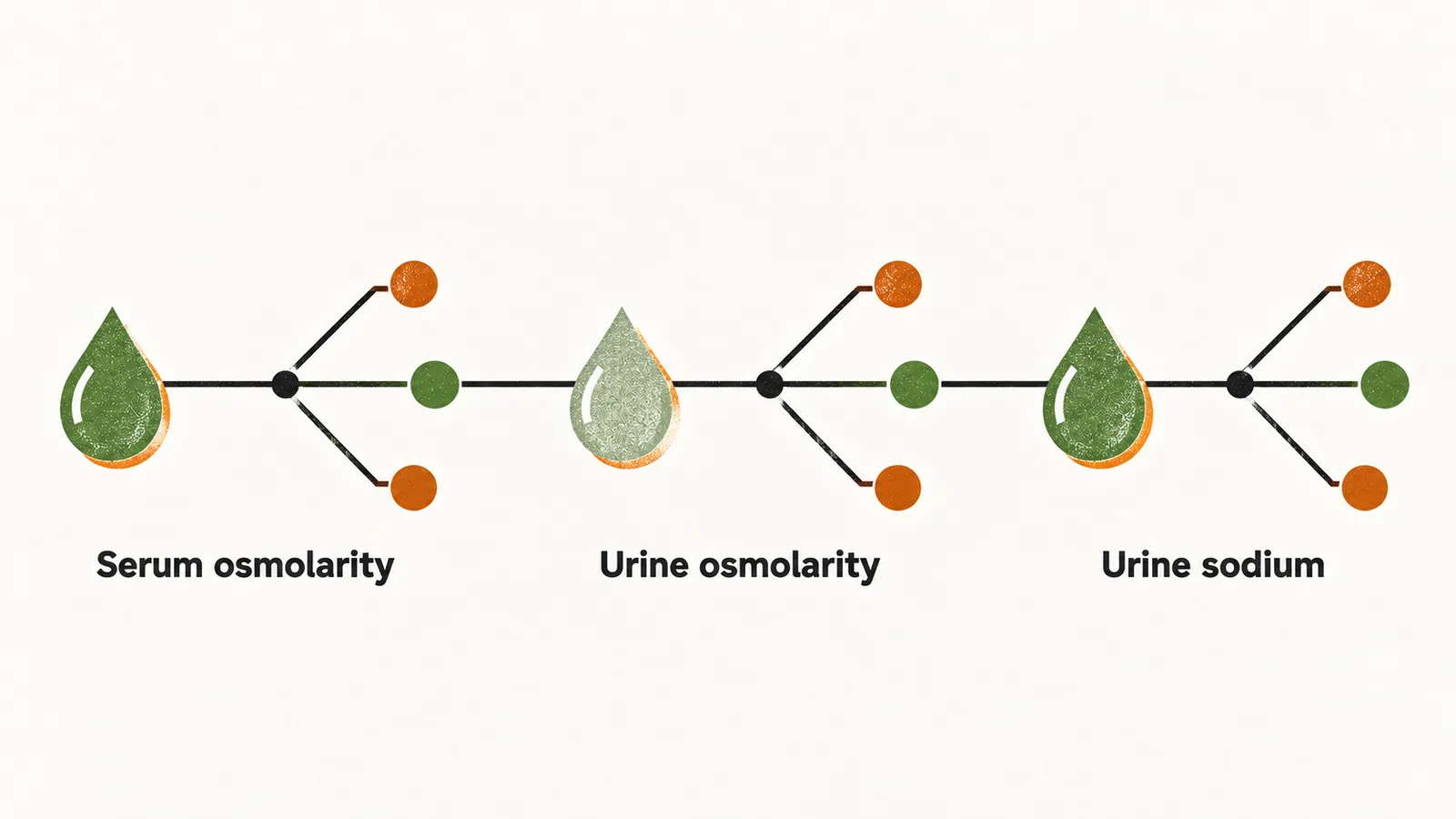
Once the bedside has suggested a group, three laboratory tests narrow the differential. Each answers one question. Serum osmolarity shows whether the low sodium reflects a truly hypotonic state. Urine osmolarity shows whether vasopressin is acting. Urine sodium shows whether the kidney is retaining sodium.
Is the serum really dilute?
The first question is whether the solution around the sodium is truly dilute. Serum osmolarity is mainly determined by sodium and its associated anions, as Osmolality, Osmolarity and Tonicity shows, so it falls in most patients with hyponatremia. Normal serum osmolarity is 275 to 290 mOsmol/kg. The value divides hyponatremia into hypotonic and non-hypotonic forms. Most hyponatremic patients are hypoosmolar, so most have the hypotonic form.
Can sodium be low when the solution is not dilute?
Yes, because a laboratory value can read low when nothing is dilute. Sodium can be low with normal osmolarity, as in severe hyperproteinemia or hyperlipidemia. The reason is that sodium lives in the water part of plasma. The protein or lipid takes up space and shrinks that water fraction, so the same sodium is measured in a smaller plasma water fraction and reads low. The true concentration is normal.
Raised osmolarity points the other way, to another solute in the plasma, such as glucose in hyperglycemia. Such a solute stays outside the cells and pulls water out of them.
What does urine osmolality show?
If the serum is truly dilute, the next question is whether ADH is acting. Urine osmolarity tests the normal response to hyponatremia, which is a reduction in AVP release that dilutes the urine as much as possible. The kidney has done that when the urine is dilute, so an appropriate response gives a urine osmolarity less than 100 mOsmol/kg. A urine osmolarity over 100 reads the same threshold the other way: it indicates excessive action or production of AVP.
Why can the same urine mean different things?
The urine has to be read against the volume state that the examination found. ADH is also released to protect volume, as the causes note describes, so a concentrated urine in a hypovolemic patient is an expected response. The same urine results carry a different meaning in a hypovolemic patient than in a euvolemic one.
What does urine sodium add?
Urine sodium asks whether the kidney is retaining sodium, and so it separates the causes that reduce effective arterial volume from those that do not. A urine sodium less than 30 mmol/L suggests that the kidneys retain sodium because effective arterial volume is low. That occurs in hypovolemia, heart failure or cirrhosis. A urine sodium above 30 mmol/L in a patient who is not fluid-overloaded fits SIAD, because in SIAD the arteries are full and the kidney has no need to hold sodium.
A urine sodium above 30 mmol/L is not specific on its own. Diuretics, adrenal insufficiency and other causes can raise urine sodium. So one value cannot settle the group.
How is the severity judged?
With the group known, the next question is how urgent the case is. The approach depends on the severity of the manifestation at presentation. Severity here means symptoms rather than the number alone. Severe symptoms, namely vomiting, seizures, coma or cardiorespiratory distress, are a medical emergency.
How is severe hyponatremia treated?
Severe symptoms are treated with hypertonic saline: 3% saline, 150 mL over 20 minutes. Sodium holds water in the extracellular space, so a concentrated sodium dose raises plasma sodium quickly. The saline is repeated until the symptoms improve or the sodium has risen by about 5 mmol/L. The rounds stop when the symptoms ease, because that is the aim.
Why is the rise in sodium capped?
Treatment raises sodium, so the next question is how fast it can safely rise. Correction is capped to avoid injury. In chronic hyponatremia the sodium should not rise by more than 10 mmol/L in the first 24 hours, and not by more than 8 mmol/L in each subsequent 24-hour period. A faster rise risks osmotic demyelination, because the brain has adapted to the low sodium over days and a fast rise outpaces that adaptation.
What is done about the cause?
Correction alone does not finish the job, so the cause is treated in parallel. If the cause is left alone, it keeps driving water retention.
- The offending drug is stopped, because a drug that raises ADH keeps water held, and stopping it removes that drive.
- Fluid is restricted in SIAD, because with ADH acting the kidney cannot clear water, so intake must come down.
- Volume and salt are replaced in hypovolemia, because restoring volume removes the signal that keeps ADH on.
When can correction be left alone?
Chronic hyponatremia, even if very profound, is tolerated well by the body, because it has had time to adapt. So in asymptomatic chronically hyponatremic patients, correction may be skipped at diagnosis, before the cause is determined. How do the history, the tests and the severity fit together into one plan when the picture is not clean?