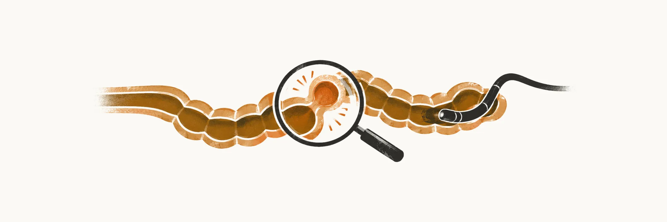
Diverticulitis is inflammation of a diverticulum, and imaging carries its diagnosis, while colonoscopy is reserved for specific problems because of its risk during an acute attack.
Colonoscopy
Colonoscopy is contraindicated in patients who are suspected of having diverticulitis, because the chance of perforation is high in these patients. The indications for performing colonoscopy in patients suspected of diverticular disease are therefore limited to defining the source of lower gastrointestinal bleeding, treating diverticular bleeding, and excluding cancer.
When colonoscopy follows an episode of diverticulitis, it should be delayed until the acute inflammation has settled — 6–8 weeks, or until the acute symptoms have resolved, whichever is longer — so that the risk of perforation is lower and the mucosa can be inspected. It is brought forward only when warning features such as weight loss or a change in stool calibre raise the possibility of cancer.
Imaging
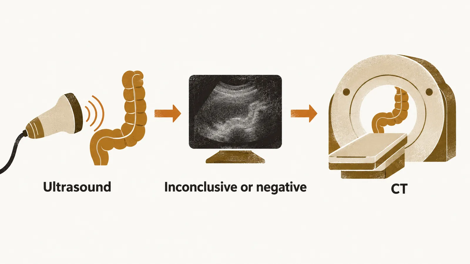
Ultrasound of the sigmoid colon by an experienced examiner and contrast-enhanced CT are both accurate, and either may come first in practice. Guidelines favour contrast-enhanced CT because it also grades severity and shows complications, while ultrasound has the advantage of avoiding radiation; when ultrasound is inconclusive or negative, CT follows. CT has a sensitivity of about 94–95% and a specificity of about 96–99% for the diagnosis of colonic diverticulitis. CT with contrast uses water-soluble oral, rectal and intravenous contrast agents.
The main findings on abdominal CT with contrast in patients with diverticulitis are outpouching of the colonic wall — normally the outer wall of the colon is smooth, so outpouching indicates diverticulitis — thickening of the colonic wall, stranding of the peri-colic fat (a streaky haze in the fat around the colon), peri-colic fluid accumulation, an abscess, and extravasated contrast agent, meaning contrast that has leaked out of the bowel.
Alongside these studies, a plain radiograph of the abdomen should be performed in any patient presenting with acute abdominal pain, to exclude perforation and obstruction.
Differential diagnosis
Imaging also has to separate diverticulitis from the conditions that resemble it, and the differential diagnosis is extensive. Right-sided diverticulitis is most often misdiagnosed as appendicitis. Inflammatory bowel disease (IBD) and colorectal cancer are the other colonic conditions to exclude, and cancer is one reason colonoscopy is arranged once the attack has settled. Three gynaecological conditions can also produce a similar picture: pelvic inflammatory disease, ovarian cyst rupture and ovarian torsion.
Once the diagnosis is made and complications are looked for, the next decision is how to treat, and that depends on whether the disease is uncomplicated or complicated.