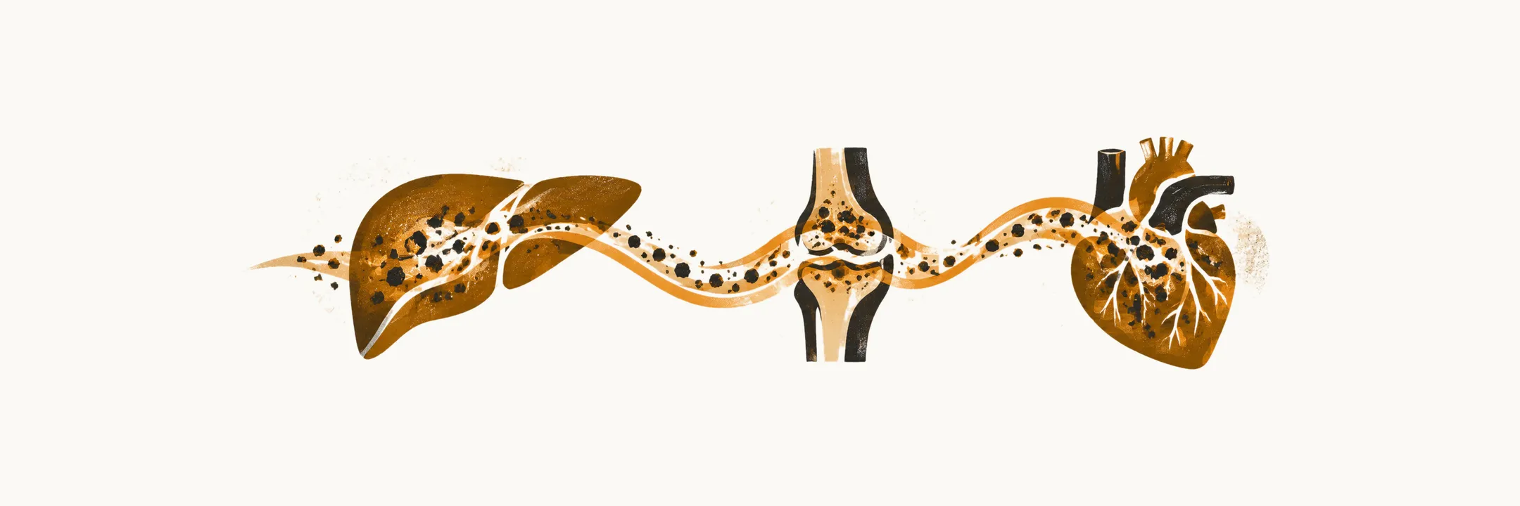
Hereditary hemochromatosis (HH) usually develops silently, and when symptoms do appear they come from iron deposited in a few characteristic organs: the liver, pancreas, pituitary gland, heart and joints.
How it presents
Most patients with HFE-related HH (the form caused by mutations in the HFE gene) come to medical attention in the asymptomatic phase of the disease, for example when relatives of a symptomatic homozygous patient are tested as part of family screening.
In patients who do develop symptoms, the most common presentations of HFE-related HH are weakness and lethargy, abdominal pain (mostly vague pain in the right upper quadrant) and arthralgia, or joint pain. The joint pain of hemochromatosis has a characteristic distribution: it typically affects the second and third metacarpophalangeal joints, and calcium pyrophosphate crystals can be deposited in the cartilage (chondrocalcinosis).
Beyond these general symptoms, the endocrine and liver effects follow the organs that hold the iron.
Loss of libido and potency in men has two mechanisms: testicular failure from iron overload of the gland, and a lack of gonadotropin (the pituitary hormone that drives the testes) from iron damage to the pituitary gland. Hypothyroidism from iron overload of the pituitary has also been reported, similar to the gonadotropin effect.
Liver-related manifestations include hepatomegaly (an enlarged liver), and less commonly ascites, edema, jaundice and splenomegaly.
Complications
The end-stage complications, which were very frequent in the past, are diabetes mellitus, cirrhosis, skin hyperpigmentation, hepatomegaly and heart failure. During the past years, thanks to the screening tests and the earlier diagnosis of HFE-related HH, the total incidence of these complications has fallen dramatically and many are now prevented.
Age, sex and penetrance
Most patients who are symptomatic are diagnosed between 40 and 50 years of age, and homozygosity for the C282Y mutation is found equally in men and women. Clinical penetrance, the proportion of people with the genotype who develop disease, is however lower in women than in men, because blood loss during childbirth and the menstrual cycle removes iron. Before screening was widespread, women presented about ten years later than men and were greatly outnumbered among symptomatic cases; with screening the two sexes are identified at similar rates, but the proportion of C282Y homozygous women who develop definite disease remains much lower than in men.