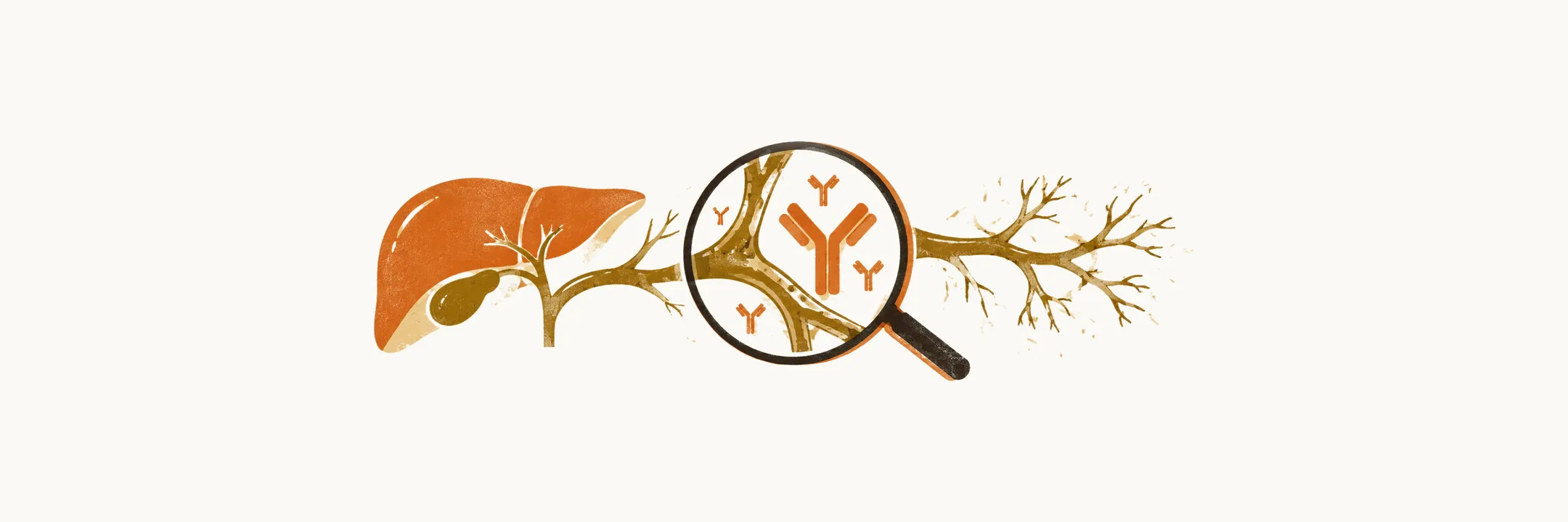
The diagnosis of primary biliary cholangitis (PBC) usually rests on the liver tests and the autoantibody profile rather than on a liver biopsy.
Diagnosis
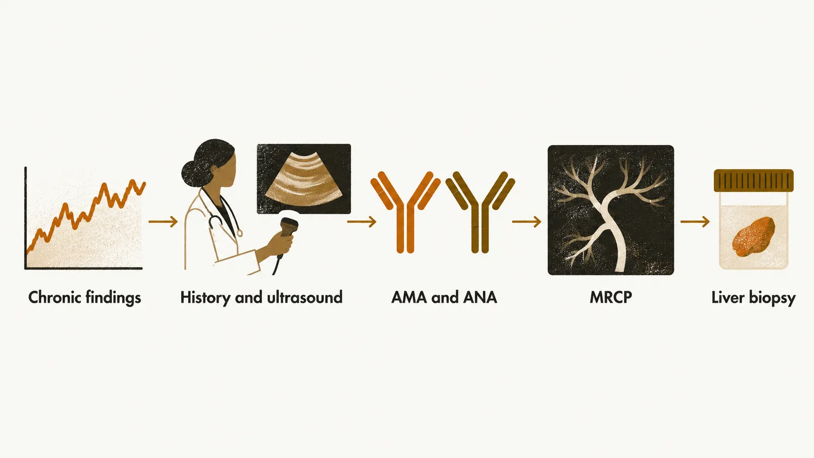
PBC should be suspected when a patient has cholestatic laboratory or clinical findings (signs of impaired bile flow) that have run a chronic course, for longer than 6 months. When that pattern is present, the workup proceeds in steps, each taken only if the one before does not settle the question:
- History, physical examination and abdominal ultrasound. Ultrasound is done first to look for a dilated biliary tract, which would point to gallstones or another obstruction rather than to PBC.
- Testing for anti-mitochondrial antibody (AMA) and antinuclear antibody (ANA).
- If these are negative, extended imaging such as magnetic resonance cholangiopancreatography (MRCP), which images the bile ducts.
- If this is also unrevealing, liver biopsy.
The diagnosis is confirmed when at least 2 of the following 3 criteria are met in the patient:
- Chronic elevation of the liver tests, mainly alkaline phosphatase (ALP) at least 1.5 times the upper limit of normal. Almost all patients with PBC have raised ALP and gamma-glutamyl transferase (GGT), the two enzyme tests that rise with cholestasis. ALT and AST, the aminotransferases, are usually only mildly raised, less than 3 times the upper limit of normal, and a marked elevation above 5 times the upper limit is unusual and raises the possibility of an overlap syndrome. The serum bilirubin is normal in the early stages and rises slowly with progression, and serum immunoglobulins, especially IgM, are increased.
- AMA positivity at a titre above 1:40. The most common technique for detecting AMA is indirect immunofluorescence (IIF); ELISA for the M2 subtype is more sensitive and specific, about 90% sensitive and 96% specific, and is recommended when IIF is negative. Other autoantibodies are often present: rheumatoid factor (RF) in about 70%, smooth-muscle antibody (SMA) in about 66%, ANA in about 50%, and anti-thyroid antibodies in about 41%.
- Histological findings consistent with PBC. One of the earliest changes is loss of the canals of Hering, and the most common, and often the only, diagnostic clue is ductopenia, loss of bile ducts in more than 50% of the portal tracts.
The histological changes can be graded with the Ludwig staging system, which has four stages:
- stage 1 — focal lesions only around the bile ducts of the portal tract, known as florid duct lesions
- stage 2 — expansion of the lesion from the portal tract into the parenchyma, that is, interface hepatitis
- stage 3 — fibrosis and scarring
- stage 4 — a cirrhotic liver biopsy with fibrous septa and regenerative nodules
Liver biopsy is not generally required, because most patients are confirmed by serology and biochemistry and 2 of the 3 criteria are enough. When the serological and biochemical evidence is not sufficient, a liver biopsy is performed.
Once the diagnosis is established, the question becomes how to treat the disease, and how to judge whether treatment is working.