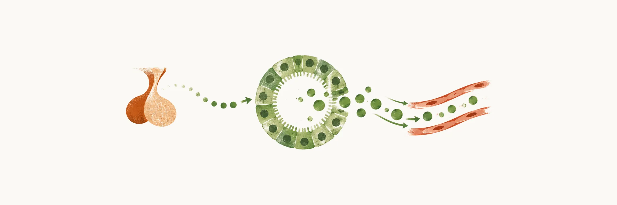
The thyroid gland makes two hormones, thyroxine (T4) and triiodothyronine (T3). It does not set its own output. Its activity is set by thyroid-stimulating hormone (TSH), which the anterior pituitary gland releases under the influence of hypothalamic thyrotropin-releasing hormone (TRH). Thyroid hormone then feeds back on the pituitary and hypothalamus to turn that signal down. Where the gland lies, how it is built and how TSH controls it explain how the hormones are made and released.
Where does the gland sit?
The gland lies in the front of the neck, anterior to the trachea, between the cricoid cartilage and the suprasternal angle. In adults it normally weighs about 12–20 grams. It is soft in consistency.
What lies beside it?
The gland lies in front of the windpipe, so the next question is what else sits close to it. Four parathyroid glands sit posterior to the poles of the gland, the inferior pair at the lower poles and the superior pair near the upper poles.
The recurrent laryngeal nerve runs close to the lateral borders of the gland. It reaches close to the trachea at the lower pole and usually lies deep to the parathyroid gland. The nerve supplies most muscles of the larynx, and injury to it causes vocal cord paralysis. Work on the gland is also work near the nerve, and that is why protecting the recurrent laryngeal nerve during thyroid surgery matters.
What is the gland built from?
Those are the gland’s neighbours. The gland itself is made of many thyroid follicles, so what is inside one? Each follicle is a ring of follicular cells around a lumen filled with thyroglobulin. Thyroglobulin is the protein precursor of the hormones. It is an inactive storage form of thyroid hormone, so the follicle holds its hormone in the protein, outside the cell. It is also the molecular template on which T4 and T3 are made, so the stored protein is both the store and the template.
The gland is highly vascular. The follicles sit in a bed of capillaries, and these capillaries carry the hormone away to where it acts.
How does the gland get its follicles and its switches?
The follicle and its stored protein are in place before any hormone is made, so how does the gland get built? The gland first produces thyroid hormone during the 11th week of gestation.
The C cells are classically said to come from neural crest cells. Recent mouse lineage tracing argues that they also derive from pharyngeal endoderm, so the origin of the C cells is debated.
Development of the gland depends on the transcription factors TTF-1, TTF-2 and PAX-8. They are expressed in the early phases of thyroid organogenesis. They work by switching on the genes for the gland’s own machinery: thyroglobulin (Tg), thyroid peroxidase (TPO), TSH and the TSH receptor (TSH-R).
This explains a cause of congenital hypothyroidism. A mutation in the gene for one of these transcription factors, or in one of the genes they control, can cause congenital hypothyroidism. Such cases are rare.
What does TSH do at the follicle?
When the body needs more hormone, TSH acts on the basolateral surface of the follicular cells. The main site of TSH receptor expression is this membrane, so the signal arrives from the blood.
Under TSH the follicular cells take thyroglobulin back in, so the stored protein is pulled into the cell. Inside the cell, thyroglobulin is broken down to release the hormone. The follicular cells then pass the hormone into the bloodstream, through the capillaries around the follicles. TSH sets this whole process going, so knowing where it comes from and how it is regulated helps.
Where does TSH come from, and what is it made of?
TSH acts on the follicle, so where is it made? The main controller of the axis is TSH, and it is made by the thyrotropic cells of the anterior pituitary gland. The hypothalamus controls the anterior pituitary, and the pituitary’s TSH is what reaches the gland, so the pituitary is the middle link of the chain. TRH reaches the anterior pituitary through the hypothalamic-hypophyseal portal system, so the signal travels in blood. The cells that make TSH are the same cells TRH acts on, so the TRH signal meets them directly.
Like other glycoprotein hormones, TSH has two subunits, alpha and beta. The alpha subunit is shared with hCG, LH and FSH, so it cannot by itself tell these hormones apart. The beta subunit is unique to TSH. It is the hormone-specific part, and it gives TSH its specificity.
What turns TSH down?
TSH is made in the pituitary, so what tells the pituitary to slow down? TSH secretion is restrained mainly by negative feedback from thyroid hormones. They act on the pituitary and hypothalamus through the TRβ-2 receptor, a subtype of thyroid hormone receptor. Feedback through TRβ-2 suppresses both TSH and TRH, so one receptor lowers both steps of the chain. In a mouse study, TRβ-2 was found restricted to the thyrotrophs and the TRH neurons of the paraventricular hypothalamus, and this restricted pattern is suggested to make it the mediator of T3 negative feedback on TSH and TRH.
Thyroid hormone negative feedback is the dominant regulator of TSH production. A few other signals can lower TSH, but only when present in pharmacological dose: dopamine, somatostatin and glucocorticoids.
How steady is TSH in the blood?
Feedback sets TSH over time, so how well does a single TSH value show what the gland is doing? TSH has a long half-life for a pituitary hormone, about 50 minutes. Because of this, the TSH blood level is steady and tracks the hormone level closely. That makes TSH a very good laboratory target for disorders of the thyroid axis.
Where does the signal go next?
Once TSH reaches the gland, activation of the TSH receptor stimulates the follicular cells to take up iodine and to build T4 and T3 from it. How does a follicular cell take up iodine, and how does it build T4 and T3 from that iodine?