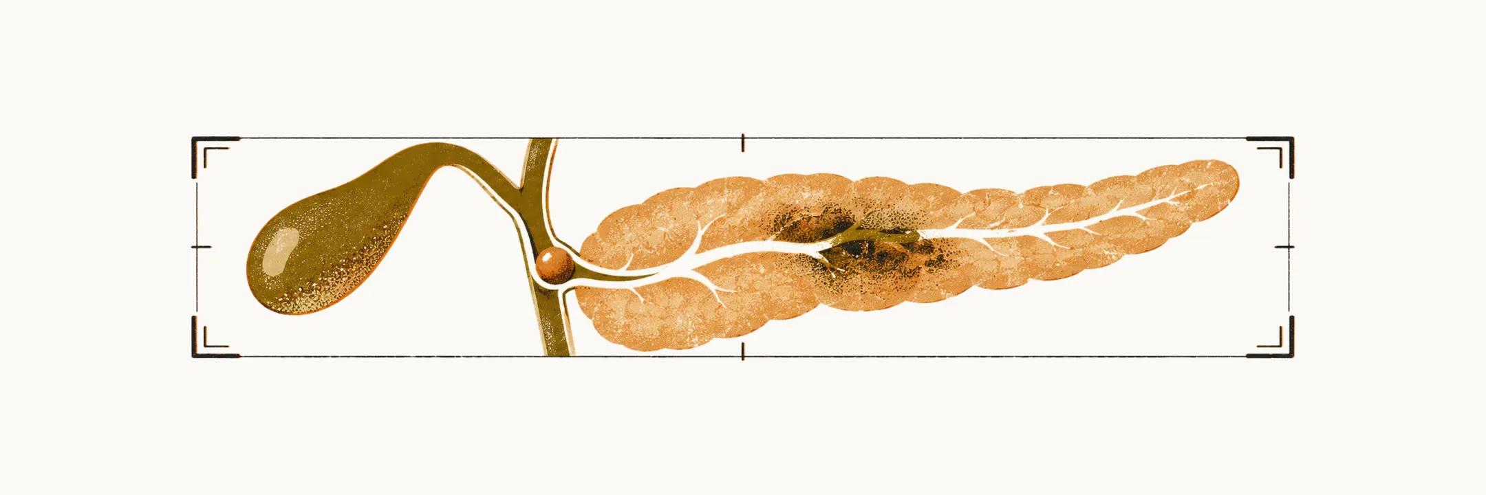
Imaging in acute pancreatitis has four jobs: finding the cause, excluding other abdominal emergencies that look similar, grading severity, and detecting complications. Each modality is chosen for what it shows best, from the early test that looks for gallstones to the scan that shows necrosis and collections.
Looking for the cause
Abdominal ultrasound is useful in the first 24 hours of hospitalisation for the detection of a gallstone or dilation of the common bile duct due to cholelithiasis, and for the detection of ascites.
Endoscopic ultrasound (EUS) is recommended in cases of idiopathic acute pancreatitis, 8–12 weeks after the attack, to detect the possible presence of microlithiasis, small ductal tumours causing obstruction, and chronic pancreatitis presenting as acute pancreatitis.
Plain radiography
Plain radiography of the abdomen is used mainly for two purposes: to exclude other causes of abdominal pain, such as bowel obstruction and perforation, and to find findings suggestive of acute pancreatitis and its complications. The findings it shows are mostly related to the complications of the course: localised ileus (bowel that has stopped moving its contents onward), generalised ileus, the colon cut-off sign (pancreatic exudate induces colon spasm, with proximal air and dilation) and ascites. Plain radiography of the chest shows respiratory complications in about 30% of patients: elevation of the hemi-diaphragm, pleural effusion and pulmonary infiltrate.
CT and MRI for severity and complications
Computed tomography (CT) is the most important diagnostic tool for acute pancreatitis and its complications. It is indicated in patients with acute pancreatitis for three main reasons: to exclude other disorders that can mimic the disease, such as mesenteric infarction; to determine severity, which is assessed on un-enhanced CT across five levels from A to E; and to confirm the presence of extra-pancreatic complications. A contrast agent guides CT in differentiating pancreatic necrosis from simple interstitial pancreatitis with normal perfusion. Gas bubbles on the scan suggest pancreatic infection as a complication. CT is used for the detection of pancreatic necrosis, peri-pancreatic inflammation and fluid accumulation, and other organ failure.
Magnetic resonance imaging (MRI) has the same usefulness as CT in showing the severity of acute pancreatitis, but is better at showing peri-pancreatic fluid collections, because it distinguishes fluid from necrotic debris more clearly.