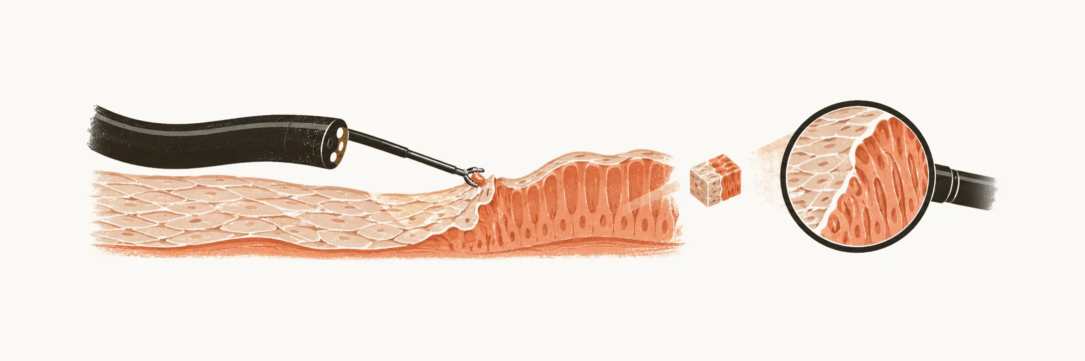
Barrett esophagus is a change in the lining of the distal esophagus, so it is diagnosed by looking at the lining and examining a sample of it. The difficult part is not finding the metaplasia but detecting dysplasia, the step toward cancer, within it.
Diagnosis
The diagnosis of Barrett esophagus is confirmed only when two findings are present together:
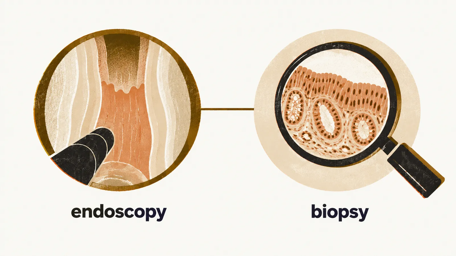
- Barrett metaplasia seen at endoscopy in at least 1 cm of the distal esophagus
- pathological confirmation of columnar cells in the endoscopic biopsy
At endoscopy the metaplastic columnar mucosa looks reddish and salmon-coloured, while the stratified squamous epithelium is pale and glossy — the change in colour marks the border between the two linings.
Almost all diagnostic criteria require intestinal-type metaplasia, the variant with mucus-secreting goblet cells (cells that are normally found in the intestine, not the esophagus), as the diagnostic finding. Some criteria also accept cardiac-type epithelium of the stomach, which is also mucus-secreting but carries a lower cancer risk.
Barrett esophagus is sub-classified by how far the metaplasia reaches above the esophagogastric junction (EGJ), the point where the esophagus joins the stomach:
- long-segment Barrett esophagus: metaplasia more than 3 cm above the EGJ
- short-segment Barrett esophagus: metaplasia less than 3 cm above the EGJ
It can also be described by the Prague C and M criteria, in which C is the circumferential extent of the metaplasia and M is its maximum extent. The two numbers fix the segment in a standard way, so the same patient is described consistently at every future endoscopy and the segment length is available to guide surveillance.
Once the segment is defined, the question is what lies within it, because the cancer risk depends on the grade of dysplasia.
Dysplasia
The hardest part of the diagnosis is detecting dysplasia in the biopsy. The distribution of dysplastic cells through the Barrett segment is random, so a biopsy can miss it, and pathologists often disagree about whether the same tissue is non-dysplastic, low-grade, or high-grade. The disagreement reflects both the pathologist’s expertise and the biology: dysplasia is patchy.
Dysplasia means disordered growth. It is defined by loss of uniformity from one cell to the next and loss of the normal architectural orientation of the tissue as a whole. Metaplasia and dysplasia are both considered pre-malignant — neither is invasive cancer — and both are potentially reversible.
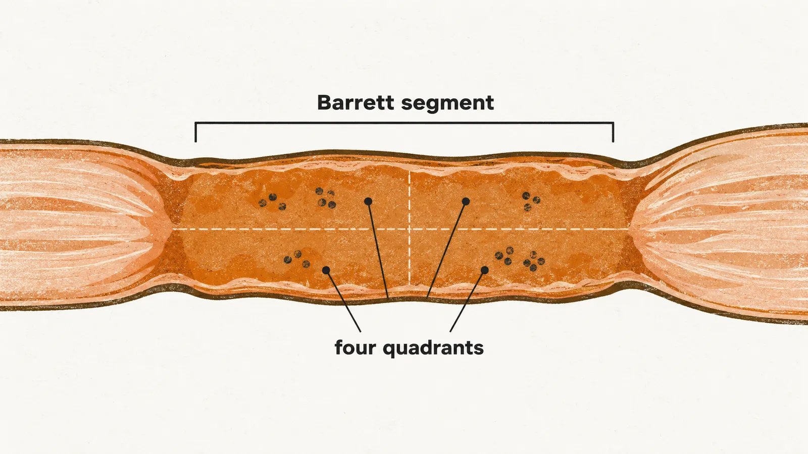
Because the abnormal cells are scattered rather than concentrated, biopsies are taken from all four quadrants of the Barrett segment rather than from one site. Current guidance calls for at least 8 biopsies in a screening examination, and the Seattle protocol for segments longer than 4 cm.
The grade of dysplasia predicts the risk of cancer. In non-dysplastic Barrett esophagus the cancer development rate is about 0.25% per year, while in high-grade dysplasia it approaches 6% per year. These rates are what make the grade decide whether acid suppression alone is enough or treatment of the dysplasia is needed.