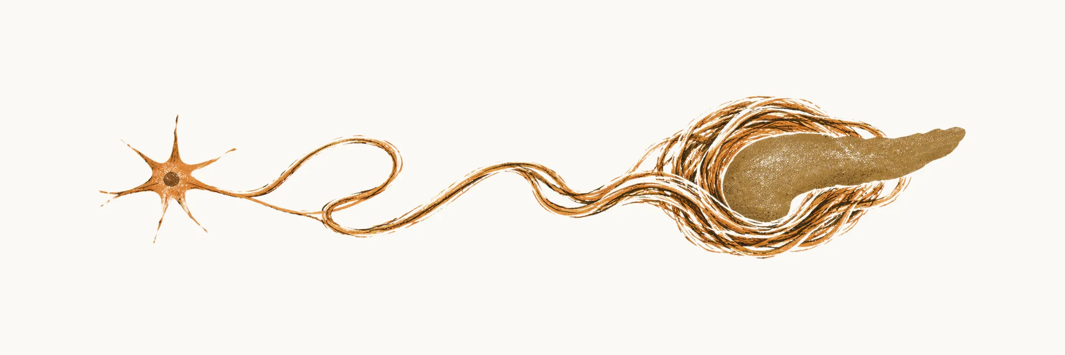
Chronic pancreatitis is driven mainly by activation of pancreatic stellate cells (PSCs). PSCs are myofibroblast-like cells (cells with features of both fibroblasts, which make connective tissue, and contractile muscle cells) of the pancreatic parenchyma, the functioning tissue of the gland. They are similar to the stellate cells of the liver, and they are the cells that build scar tissue in the gland.
When a noxious stimulus reaches the pancreas, PSCs become activated and begin to do two things at once. They release large amounts of pro-inflammatory cytokines (signalling proteins that promote inflammation), including IL-1, IL-6 and TNF-α, and they lay down fibrous tissue, mainly collagen, which is deposited in place of the normal parenchymal cells.
Pathological causes such as alcohol also act through metalloproteinase-dependent destruction of the normal collagen of the parenchyma, and this breakdown of the existing framework is what allows the gland to be remodelled into scar.
Once activated, PSCs can keep remodelling even after the original stimulus is removed. The reason is that PSCs carry TGF-β receptors, which drive further collagen production and further activation. This probably explains why the fibrosis of chronic pancreatitis continues to progress after a patient stops drinking.
A widely accepted hypothesis for how the chronic disease develops from acute attacks is the necrosis-fibrosis sequence: each episode of acute pancreatitis destroys some parenchymal cells (necrosis) and leaves a little fibrosis, and the damage accumulates over repeated episodes until the gland is chronically scarred. Repeated acute pancreatitis is therefore a route into chronic pancreatitis rather than a separate illness.
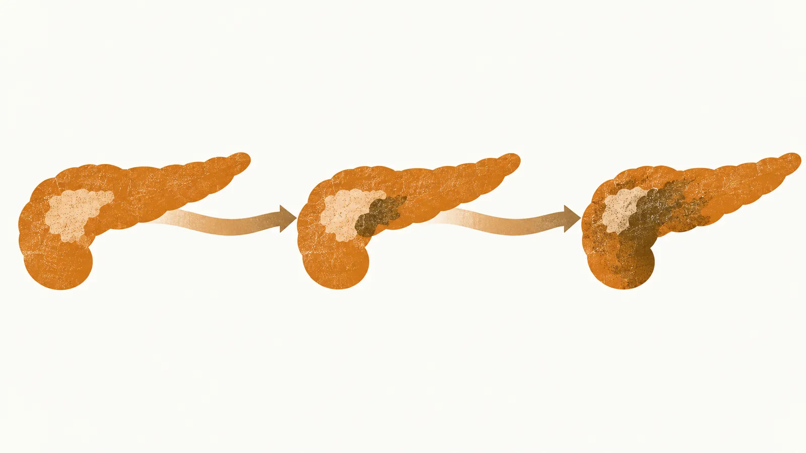
Whichever route leads there, the end result is a gland whose functioning tissue has been replaced by scar, and the clinical picture follows from that loss.