
Once the diagnosis of primary gastric adenocarcinoma is made, physical examination and imaging are used to stage the cancer, which means establishing its depth of invasion, nodal spread and distant spread, and turning those into the overall stage that guides treatment.
Imaging and endoscopic ultrasound
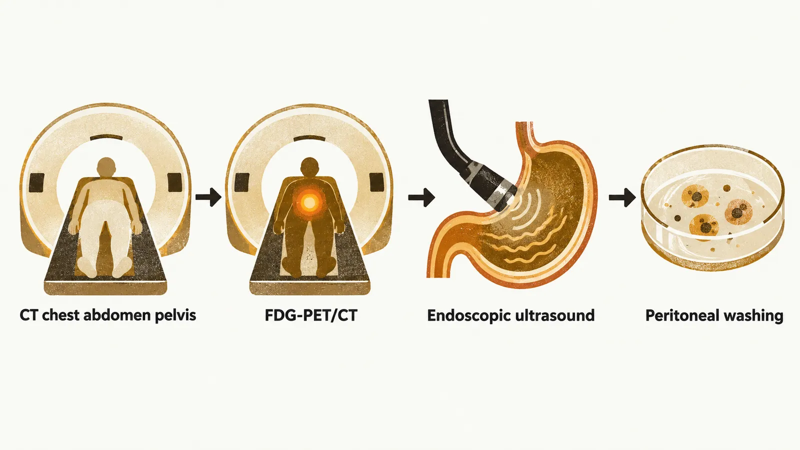
A CT scan of the chest, abdomen and pelvis should be performed. If metastasis is suspected on the CT, a biopsy should be performed to confirm stage IV disease.
When the CT is negative, FDG-PET/CT can also be performed, because it has a higher sensitivity for finding metastatic tumours. If that is also negative, the next step is endoscopic ultrasound (EUS) of the tumour itself, which is the most accurate method for assessing the depth of tumour penetration at the site of the lesion and also shows the presence of local or regional lymphadenopathy.
During the EUS evaluation, a biopsy is also taken to assess the molecular changes that direct systemic therapy: PD-L1, HER2 and MSI.
Peritoneal assessment
After that, laparoscopic investigation with peritoneal washing, in which fluid washed over the peritoneal surfaces is examined, should be considered, to find cytological changes that could be indicative of metastasis.
Naming the stage
Three staging classifications are provided for gastric adenocarcinoma: cTNM, or clinical staging; pTNM, or pathologic staging; and ypTNM, or post-neoadjuvant therapy classification. All three should be taken into account, because most patients are placed in higher stages by clinical staging, while after the pathologic classification they are mostly down-staged. The T, N and M categories they use are set out in Classification and Staging.