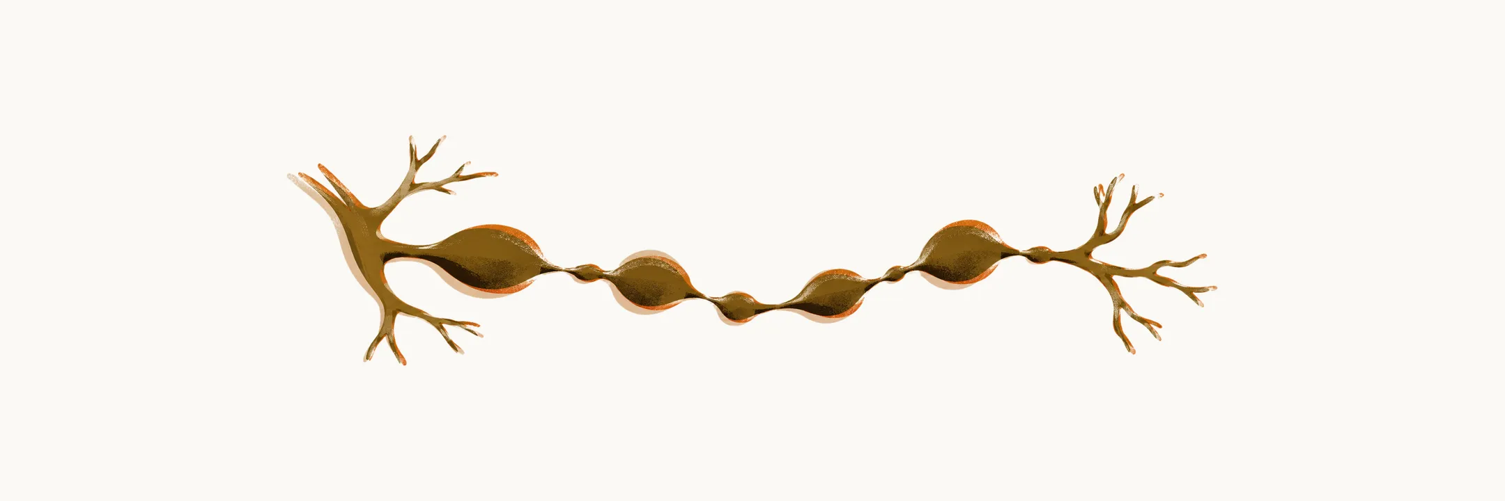
The cholangiogram — the image of the biliary tree — is what defines PSC. Its pattern of multifocal strictures and dilatations is the finding the diagnosis is built on, and it is why the disease is called sclerosing cholangitis.
How the biliary tree is imaged
The biliary tract can be imaged by three cholangiographic techniques: magnetic resonance cholangiopancreatography (MRCP), a non-invasive MRI study of the ducts; endoscopic retrograde cholangiopancreatography (ERCP), in which contrast is injected through an endoscope placed in the bile duct; and transhepatic cholangiography (THC), in which contrast is injected into the liver through the skin. MRCP is the usual first test; ERCP and THC are more invasive and are reserved for when they add information or allow treatment.
The characteristic findings
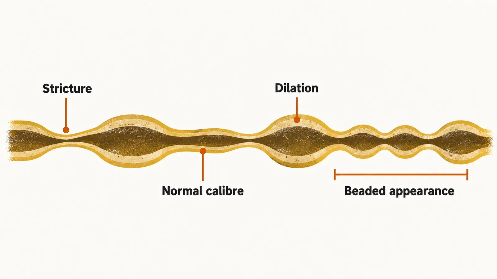
The defining feature is multifocal stricturing of the biliary tract: a stricture is a segment of duct that has narrowed, and multifocal means there are several. Areas of collapse are intercalated between areas of dilation and segments of normal calibre, which gives the ducts a beaded appearance. The dilated segments may themselves look saccular or diverticular — rounded outpouchings rather than a smooth tube. Where the ducts are dilated over a length rather than in isolated segments, the finding is called biliary ectasia.
Most cases involve both the intrahepatic and the extrahepatic bile ducts. In 15–20% of cases only the intrahepatic bile ducts are involved, and isolated extrahepatic involvement is very rare, seen in only about 6% of cases. In up to 15% of cases the cystic duct and the gallbladder are involved as well, but these structures are not easily visualised by cholangiography. Peri-portal lymphadenopathy — enlarged lymph nodes around the portal tracts — is common, seen in about 75% of cases, but it is non-specific and does not by itself indicate malignancy.
Cholangiography shows the shape of the ducts but not the tissue of the duct wall. What the wall itself looks like under the microscope is the subject of histology.