
What does ADH do, and what controls it?
The body keeps the concentration of its plasma steady, and one way it does so is by deciding how much water leaves in the urine. Antidiuretic hormone (ADH), also called arginine vasopressin (AVP), lets the kidney save water. How concentrated the blood is sets its release.
The hormone acts on the collecting duct of the kidney. This is the last part of the nephron, where the final volume and concentration of the urine are set, so it is where a hormone signal can act. ADH opens water channels in the collecting duct, so water returns to the body instead of being lost in the urine.
Where is ADH made, and how does it reach the blood?
If the blood’s concentration sets release, two things need locating: where the hormone comes from, and where the signal is read. The hormone comes first. ADH is a small peptide made by magnocellular neurons, large nerve cells. Their cell bodies sit in two hypothalamic nuclei, the supraoptic nucleus and the paraventricular nucleus.
The hormone is made in these cell bodies but delivered along their axons. The axons run down the pituitary stalk and end in the posterior pituitary. There the hormone is stored in the nerve terminals, and it is released into the bloodstream when the neurons fire, much as a neurotransmitter is released. So the posterior pituitary acts as a store, not a maker. In short, the cell bodies lie in the hypothalamus and the release site is the posterior pituitary.
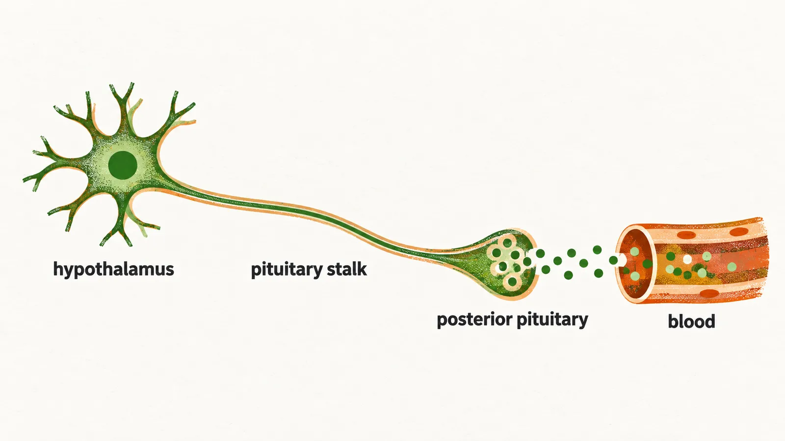
Cell bodies, axons and terminals are each needed to deliver the hormone. Damage at any point along this path, whether in the hypothalamus, the stalk or the gland, can therefore reduce ADH release. This shared pathway is why the resulting disease is called hypothalamic, or central, diabetes insipidus.
What tells the neurons when to release?
The cells that make ADH also sense the blood. The magnocellular neurons carry osmoreceptors, and osmoreceptors sense plasma osmolality, so the maker of the hormone is also its sensor. Osmolality is measured in milliosmoles per kilogram of water (mosmol/kg).
The threshold of plasma osmolality is about 280-285 mosmol/kg, and it works as a set point. Below it, ADH release is suppressed, so the kidney keeps producing dilute urine, which is what a plasma that is too dilute needs. Above it, ADH release increases and the urine is concentrated, which saves water when the plasma is too concentrated.
Thirst is regulated in parallel, at a similar threshold. Thirst is the behavioural arm and ADH is the kidney arm. A healthy person uses both: they drink and they retain water to defend plasma osmolality. Both are needed because retaining water limits the loss, and water is still lost through sweat and evaporation, so drinking replaces it.
Osmolality is not the only input. Blood volume and pressure form a second control of ADH release, and it is less sensitive. The circulation reports to the system too: a fall in pressure stimulates release. A large loss of blood volume stimulates ADH release powerfully even when osmolality is normal. The reason is that retaining water is part of defending the circulation, since water kept in the body helps keep blood volume up.
What does ADH do in the kidney?
Once released, ADH has to reach the collecting duct. It travels to the kidney and binds the V2 receptor, which sits on the principal cells of the collecting duct. These cells control how much water is reabsorbed from the forming urine, so the receptor sits on the cells that do the job.
The ADH signal inserts aquaporin-2 water channels into the apical membrane of the principal cells, the membrane facing the urine. Because the channels face the forming urine, water can leave it. Water leaves the forming urine and returns to the body, leaving a small volume of concentrated urine.
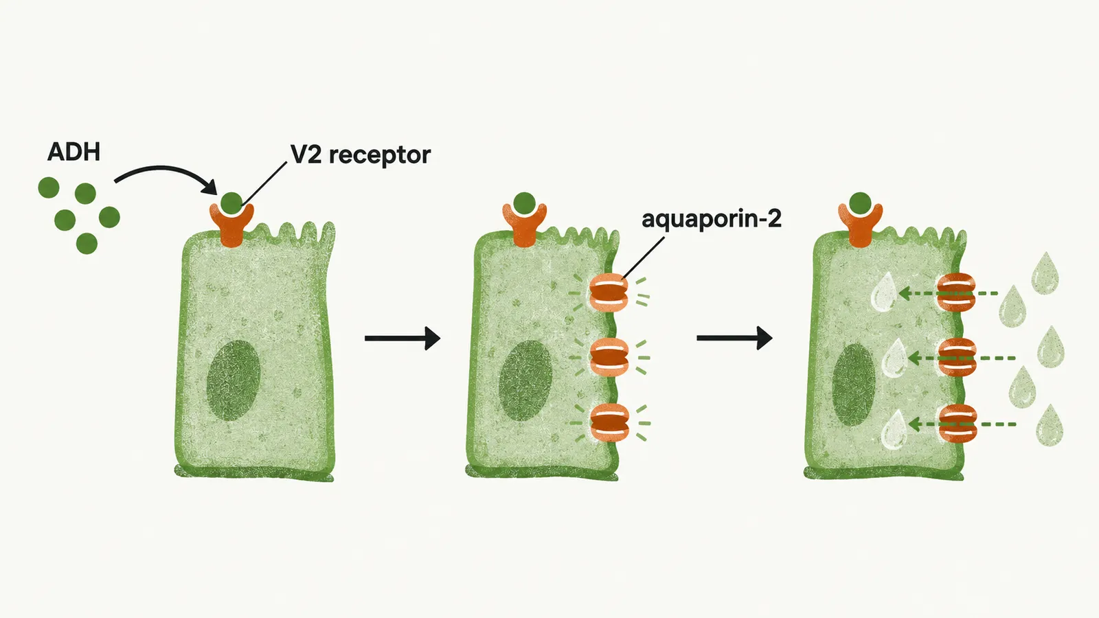
When ADH falls, the same mechanism runs backwards. The aquaporin-2 channels are withdrawn from the membrane, and the urine stays dilute.
What can go wrong?
The kidney’s response has two partners. Urine concentration depends on two things at once: enough ADH reaching the kidney, and a kidney able to respond to it.
Loss of the first, ADH, results in central diabetes insipidus. Loss of the second, the kidney’s response, results in nephrogenic diabetes insipidus. In both, the urine cannot be concentrated, and the site of failure is what names the form.
If either part fails, the urine stays dilute and its volume is large. What does that look like in a patient, and why does the plasma sodium usually stay normal?