
Calcium low enough to cause symptoms is replaced through a vein, and the patient is monitored while it is given. A calcium that is low but stable is corrected more slowly, by mouth.
Treatment has two jobs: getting calcium up, and keeping it up. The kidney is the gate for the second job. It filters calcium out of the blood and then decides how much to hand back. Normally PTH tells the kidney to keep calcium, so a rise in the blood level does not spill much into the urine. When PTH is absent, the kidney cannot hold calcium back, so every push of the blood level sends more into the urine. That is what limits the correction.
Why does severe hypocalcemia need a vein?
Hypocalcemia can be an endocrine emergency. Intravenous calcium is given without delay to severe hypocalcemia, usually a total calcium below 7.5 mg/dl. It is also given to patients with neurological manifestations: seizures, coma or altered mental status. And it is given to patients with stridor from laryngospasm or bronchospasm. In all of these the problem is the one the Clinical manifestations of hypocalcemia note describes: calcium sits outside nerve and muscle cells and steadies their membranes, so with too little calcium they fire too readily. The vein comes first because these patients cannot wait for the mouth route to act.
Which calcium is given, and how fast?
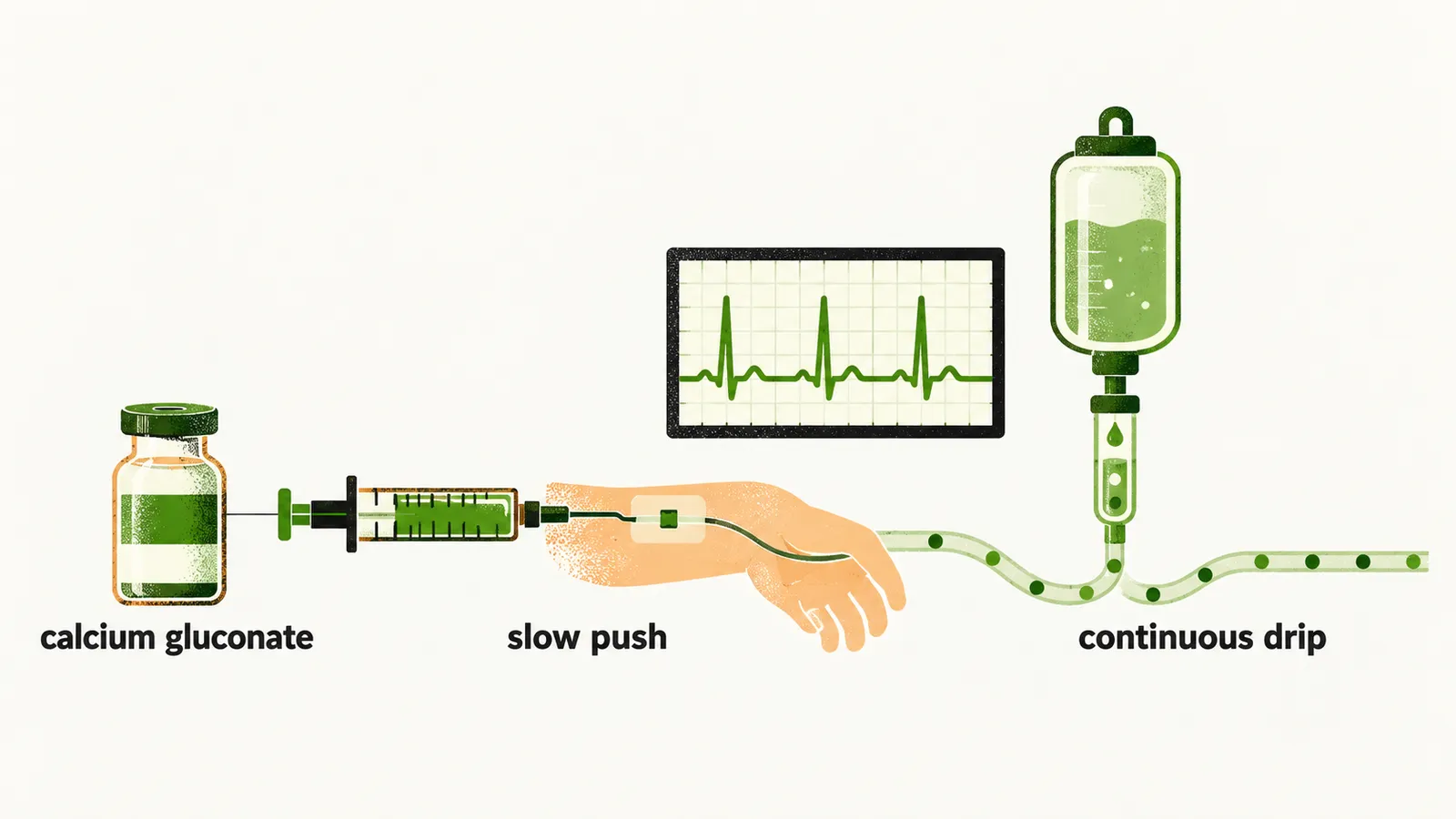
Once the vein is chosen, the drug and the rate are the next question. The usual preparation is calcium gluconate, which holds 90 mg of elemental calcium per 10 ml. It is given as slow intravenous pushes, generally one vial over 10 minutes, repeated once.
Why is the heart watched?
Calcium gluconate is given with continuous electrocardiographic monitoring. A slow push is the route for which this monitoring is recommended. Patients taking cardiac drugs, especially digoxin, are predisposed to cardiotoxicity from a calcium infusion: calcium overload raises the risk of digoxin-induced arrhythmia, so calcium given to a patient on digoxin carries the same concern. The monitoring is the reason for these cardiotoxicity precautions.
When does the drip go on, and how is it steered?
After the push, some patients are still symptomatic and oral treatment cannot act quickly enough. A continuous intravenous drip is then started to cover the time the mouth route needs. The drip rate is guided by signs, symptoms and calcium measurements. Calcium is checked every 1 to 2 hours, preferably as ionised calcium, the free, working fraction that the Diagnosis of hypocalcemia note explains.
What changes after neck surgery?
In the patient who has had recent neck surgery, the drip can be continued for longer. Parathyroid tissue may have been lost or damaged in that surgery, as the Etiologies of hypocalcemia note describes. After such surgery the oral start is often delayed. Intravenous calcium can then be continued until oral therapy is possible or has taken effect.
How is low but stable calcium corrected by mouth?
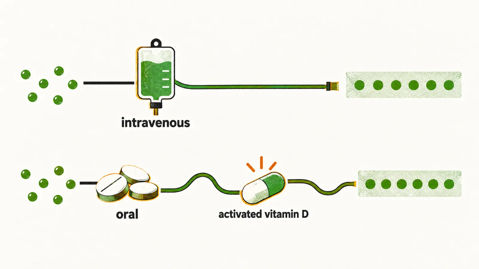
When the vein is no longer needed, the same correction moves to the mouth. A low but stable calcium is corrected more slowly, with activated vitamin D such as calcitriol. The cause of the fall is treated at the same time. Oral therapy is started as soon as practical. It uses 1 to 2 g of elemental calcium, together with a rapidly acting vitamin D such as 0.5 to 1.0 micrograms of calcitriol, in divided doses. Vitamin D has to be activated, and the kidney’s activation step needs PTH, which is why an already activated form is useful when PTH is missing.
Why calcitriol, and why split the dose?
For hypoparathyroidism, the activated analogues calcitriol or alfacalcidol are preferred. Both act rapidly and have short half-lives. Calcitriol is started at 0.25 to 0.5 micrograms once daily. Because its effect is short, it has to be given regularly, and it is uptitrated, often to twice-daily dosing, until calcium is in the target range.
Why does calcium not stay corrected?
An oral plan can still fail, and one reason is magnesium. Magnesium depletion will stop calcium staying corrected until it is addressed. Magnesium deficiency should always be considered as a contributor to hypocalcemia, and it is especially relevant in post-operative and hospitalised patients. The reason is that, as the Etiologies of hypocalcemia note explains, magnesium depletion reduces the secretion of PTH and blunts its action on the kidney. While magnesium is low, calcium and vitamin D alone will not correct the calcium level, and the calcium keeps slipping.
Why can a normal magnesium level mislead?
If magnesium is the hidden cause, the next question is how to see it. The serum magnesium can be normal or low-normal in deficiency. Magnesium is mainly an intracellular ion, so the serum level does not reflect the body’s stores. A therapeutic trial of magnesium may therefore be needed to reveal the deficiency.
How much magnesium, and for how long?
Once the deficiency is seen, it has to be replaced. Oral magnesium 200 to 300 mg daily treats mild chronic deficiency, and parenteral magnesium is used for severe hypomagnesemia. Several days of treatment are usually required to replete magnesium stores.
What limits how far calcium can be raised?
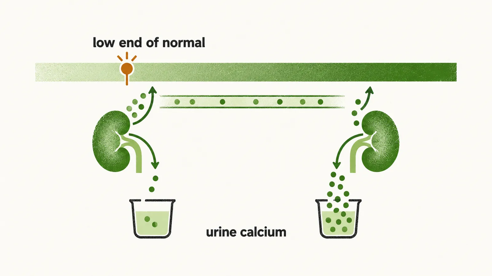
Replacement is also limited by the urine. Hypercalciuria, excess calcium in the urine, limits how far calcium can be corrected. It is especially marked in hypoparathyroidism, where the hypocalciuric effect of PTH has been lost: without PTH the kidney tubule no longer takes calcium back, so it lets it spill. Hypercalciuria predisposes to nephrolithiasis, nephrocalcinosis and renal damage. A high urine calcium is a known risk for stones, and the same spill can lead to nephrocalcinosis.
Why aim for the low end of normal?
The limit on urine calcium sets the target. The aim of chronic treatment is to keep the patient free of symptoms, with a total calcium of about 8.0 to 9.0 mg/dl, or an ionised calcium above 1.0 mmol/L. The target is the lower part of the normal reference range, deliberately. With calcium in the upper normal range there may be significant hypercalciuria, because the kidney spills more calcium for the same reason when PTH is absent.
Why can calcium and phosphate deposit in the brain and vessels?
A calcium level pushed high is not the only danger in the chronic state. A calcium times phosphate product near or above 55 mg²/dl² can cause ectopic calcification. The product can rise in hypoparathyroidism, because serum phosphate is chronically high, and a high product favours precipitation of calcium and phosphate in tissue. The deposits can appear in the brain, especially the basal ganglia, in blood vessels and in the eyes.
How is chronic hypocalcemia treated by mouth?
The chronic plan is the oral one, with its salts and its follow-up. Most causes of chronic hypocalcemia are treated with oral calcium salts together with vitamin D or an activated vitamin D analogue. Vitamin D deficiency is treated with cholecalciferol or ergocalciferol. Calcium carbonate and calcium citrate are the commonest salts. The weight of salt differs from the elemental amount: 2.5 g of calcium carbonate or 5.0 g of calcium citrate provides 1 g of elemental calcium. At follow-up, the vitamin D analogue dose is held constant, and the oral calcium is adjusted against symptoms and the calcium level.
What if hypercalciuria persists on treatment?
When urine calcium stays high on the usual doses, the question is what lowers it. A lower calcium supplement dose and a lower activated vitamin D dose are used. lower urinary calcium. A thiazide diuretic is also used. Thiazides block sodium reabsorption in the distal tubule, and they are observed to lower urinary calcium; how they do so is only proposed.
What if calcium still cannot be controlled?
If calcium or urinary calcium still cannot be controlled, recombinant human PTH (1-34) replacement is an option. It applies to chronic hypoparathyroidism, which is a lack of PTH, so it answers the missing piece itself. Does the cause of the fall change how all of this is planned?