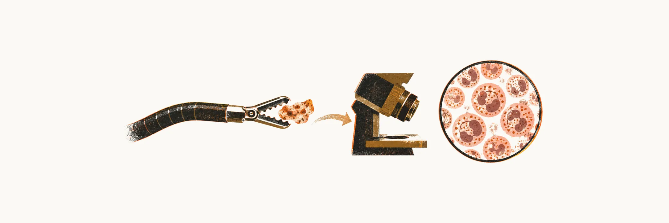
The diagnosis of eosinophilic esophagitis (EoE), a chronic eosinophil-rich inflammation of the esophagus, is made from the clinical picture together with an esophageal biopsy, and treatment then aims to keep the inflammation in check over the long term.
Diagnosis
The gold-standard diagnosis for EoE is endoscopy with pathological study of the biopsy, which is characterised by:
- 15 or more eosinophils per high-power field (HPF, the area seen at high magnification under the microscope), or about 60 eosinophils per mm²
- dilated intercellular space
- inflammation and fibrosis in the lamina propria (the connective tissue layer beneath the epithelium)
It is not adequate to make a diagnosis of EoE where there are isolated eosinophils in the biopsy without any clinical signs and symptoms.
In 2017 a structured histologic scoring system for EoE was established that grades 8 histological features and adds detail beyond the peak eosinophil count:
- eosinophil density
- basal zone hyperplasia
- eosinophil abscesses
- eosinophil surface layering
- dilated intercellular space
- surface epithelial alteration
- dyskeratotic epithelial cells
- lamina propria fibrosis
One important task during diagnosis is separating EoE from gastroesophageal reflux disease (GERD), which can be done using the number and distribution of eosinophils in the biopsy:
- fewer than 7 eosinophils per HPF points to GERD
- between 7 and 15 eosinophils per HPF points to a combination of GERD and food allergy
- more than 15 eosinophils per HPF points to EoE
- eosinophils only in the distal portion points to GERD
- eosinophils in both the proximal and distal esophagus points to EoE
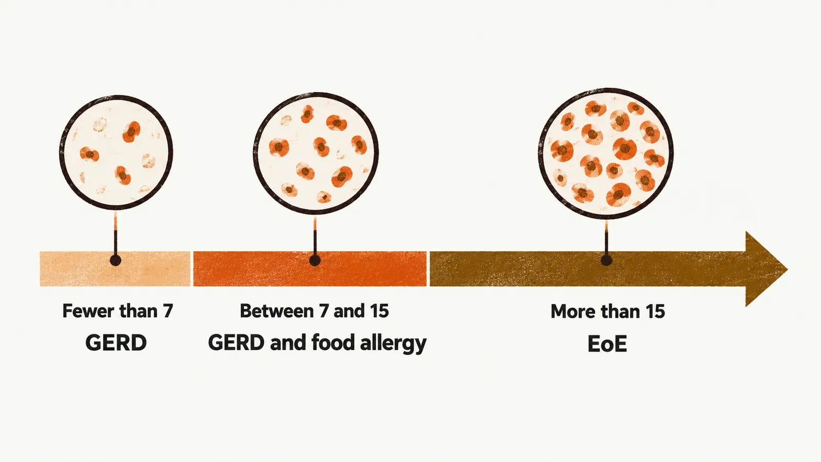
No single count or distribution is completely reliable on its own, so the diagnosis also requires a clinical assessment of the other causes of esophageal eosinophilia.
Treatment
Treatment of EoE, like the treatment of asthma, needs to be chronic and life-long: stopping it usually allows the inflammation to return. Four approaches are used to suppress it. Proton pump inhibitors are one. Swallowed topical glucocorticosteroids such as budesonide or fluticasone act directly on the esophageal lining, and their main adverse effect is local fungal or viral infection. Elimination diets run from an elemental diet to the empiric removal of common foods. The fourth is the biologic dupilumab, which is directed against the IL-4 receptor.
Complications and involvement beyond the esophagus
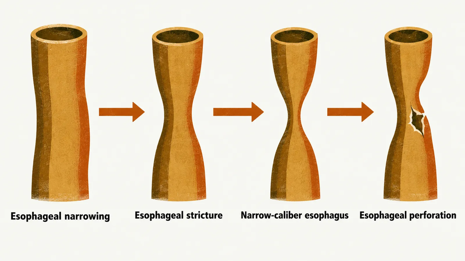
If EoE is left untreated, progressive esophageal dysfunction and scarring can lead to esophageal narrowing, esophageal stricture, a narrow-caliber esophagus and esophageal perforation. Endoscopic dilation relieves a stricture but does not treat the inflammation underneath it.
EoE is not always confined to the esophagus, because the non-esophageal EGIDs can occur alongside it. Gastric and duodenal biopsies are therefore taken when symptoms or endoscopic findings point to involvement, rather than as routine surveillance of the whole GI tract.