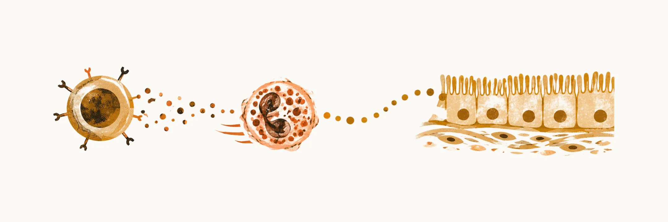
Eosinophilic gastrointestinal disorders arise from an immune response that recruits eosinophils, a type of white blood cell normally scarce in the gut wall, into the wall of the gastrointestinal tract and keeps them there.
EGIDs have properties that fall between pure IgE-related food allergy and cellular-mediated hypersensitivity disorders, so they do not fit neatly into either category.
Genetic and environmental factors
The pathophysiology of the EGIDs is related to an interplay between genetic factors, injurious agents, and environmental factors such as diet. Eosinophilic esophagitis (EoE) clusters in families: about 2% of first-degree relatives of an affected person also have EoE, and the recurrence risk in relatives is raised roughly 10- to 64-fold, with brothers at the highest end. That pattern, a raised but far from certain familial risk with a male predominance and no clean Mendelian ratio, points to a complex polygenic inheritance shaped strongly by shared environment rather than to a single causative gene.
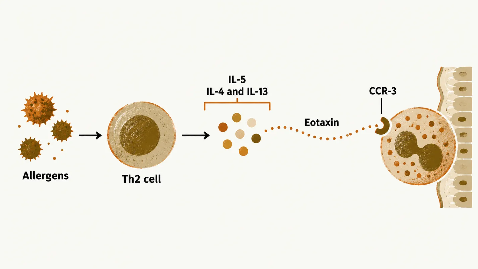
Within that susceptibility, several factors contribute to the inflammatory cascade. Allergens activate Th2-polarised T cells, helper T cells that drive allergic-type inflammation. These cells release cytokines (signalling proteins) such as IL-5 and IL-13. Chemokines (signalling proteins that guide cells to a tissue), micro-RNA (small regulatory RNA molecules), a loss of epithelial barrier function, and an altered balance between proteases and their inhibitors also contribute.
Eosinophil recruitment and its mediators
Normally eosinophils home to the GI tract, lymph nodes and spleen. In patients with EGIDs this recruitment increases and extends to regions of the GI tract that are not normally recruited, such as the Peyer patches (lymphoid tissue in the wall) of the small bowel. The recruited eosinophils are driven by the cytokines IL-5, IL-4 and IL-13, which are made by the Th2 cells. IL-5 is the most important signal for the development and persistence of eosinophils in the GI tract, while IL-4 and IL-13 also act even in the absence of IL-5. Studies show that reducing IL-5, IL-4 and IL-13 produces some improvement in patients with EGIDs, especially EoE.
Recruitment into the tissue is guided by chemokines, and one of the main ones is eotaxin, recognised by the eosinophil receptor CCR-3. The Th2 cytokines, eotaxin and CCR-3 are all possible diagnostic, treatment and pathological targets.
Eosinophil accumulation is not proof of an EGID
Accumulation of a high number of eosinophils in the GI tract is not always indicative of EGIDs. In several different diseases, such as gastroesophageal reflux disease (GERD) or inflammatory bowel disease (IBD), eosinophils accumulate in the GI tract, but the inflammation is not eosinophil-dominant. The diagnosis therefore rests on eosinophil-rich inflammation without a secondary cause, together with GI symptoms.
Eosinophil numbers in the blood vary between patients. In more than 50% of the cases of EGIDs the disease is isolated, with no peripheral eosinophilia (raised eosinophils in the blood), while in some patients, especially those with eosinophilic gastritis, there is a high level of peripheral eosinophils that can be considered as part of a hypereosinophilic syndrome, or HES.