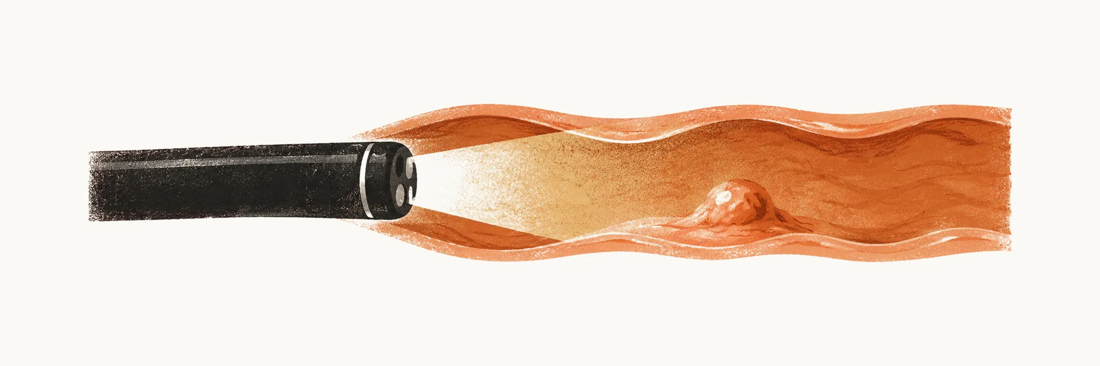
Diagnosing an esophageal disorder means choosing among several tests, each of which answers a different question. Endoscopy examines the mucosa directly, contrast radiography shows structure and function, manometry measures contraction, and reflux testing measures acid exposure.
Endoscopy
Endoscopy, also called esophagogastroduodenoscopy (EGD), is the most useful diagnostic investigation for the proximal gastrointestinal tract (the esophagus, stomach and duodenum). It consists of a tube with a high-resolution color camera and an instrumentation channel that allows biopsy, drug delivery, balloon dilation, and more. Its advantages over barium radiography of the esophagus are greater sensitivity for mucosal lesions and for diagnoses that depend on the color of the lesion, such as Barrett esophagus (replacement of the normal esophageal lining by a different type of epithelium) and vascular lesions; the ability to take a biopsy; and the ability to dilate a stricture during the same examination.
Contrast radiography
Endoscopy has not made conventional radiography of the esophagus obsolete, because some diagnoses are still made better by radiography, and either a positive or a negative barium study is usually followed by endoscopy for biopsy or confirmation. Contrast radiography of the esophagus, stomach, and duodenum can show reflux of the contrast medium, a hiatal hernia, mucosal granularity, mucosal erosions, mucosal ulceration, and strictures; for strictures, barium radiography is more sensitive than endoscopy. It is not only static imaging: dynamic rapid-sequence or video fluoroscopic imaging can also be performed, and it is more useful for detecting disorders of the cricopharyngeus muscle.
Endoscopic ultrasound
Endoscopic ultrasound (EUS) combines endoscopy with an ultrasound transducer. It is used mostly to evaluate dysplasia in Barrett esophagus and to assess submucosal lesions.
Esophageal manometry
Esophageal manometry, or motility testing, uses a catheter of pressure-sensing transducers to measure the change in pressure at different points along the esophagus after a swallow. It is used mostly to diagnose or assess achalasia, in which the lower esophageal sphincter fails to relax; diffuse esophageal spasm (DES); and the integrity of peristalsis before surgery for reflux disease. It can be combined with impedance sensors, which record the backward or forward movement of the bolus.
Manometry tracings are interpreted with the Chicago classification version 4.0, which rests on three measurements. Distal latency (DL) is the interval from the start of relaxation of the upper esophageal sphincter to the contractile deceleration point, and a short distal latency defines a premature contraction. Integrated relaxation pressure (IRP) is the residual integrated pressure across the lower esophageal sphincter during swallow-induced relaxation. The distal contractile integral (DCI) summarizes the vigor of the distal contraction.
Reflux testing
In gastroesophageal reflux disease (GERD) the esophagus can look normal, so endoscopy and radiography do not always settle the question: in non-erosive reflux disease endoscopy shows no sign of inflammation, yet the patient still has symptoms, because the disorder is one of over-exposure of the esophagus to gastric contents rather than of visible mucosal injury. Reflux testing measures the pH of the esophagus continuously for 24 hours, or up to 96 hours with a wireless device, and reports the acid exposure time: the proportion of the monitored period during which the pH is below 4. A distal acid exposure time below 4% is normal, above 6% is conclusively abnormal, and 4–6% is inconclusive (the Lyon Consensus).