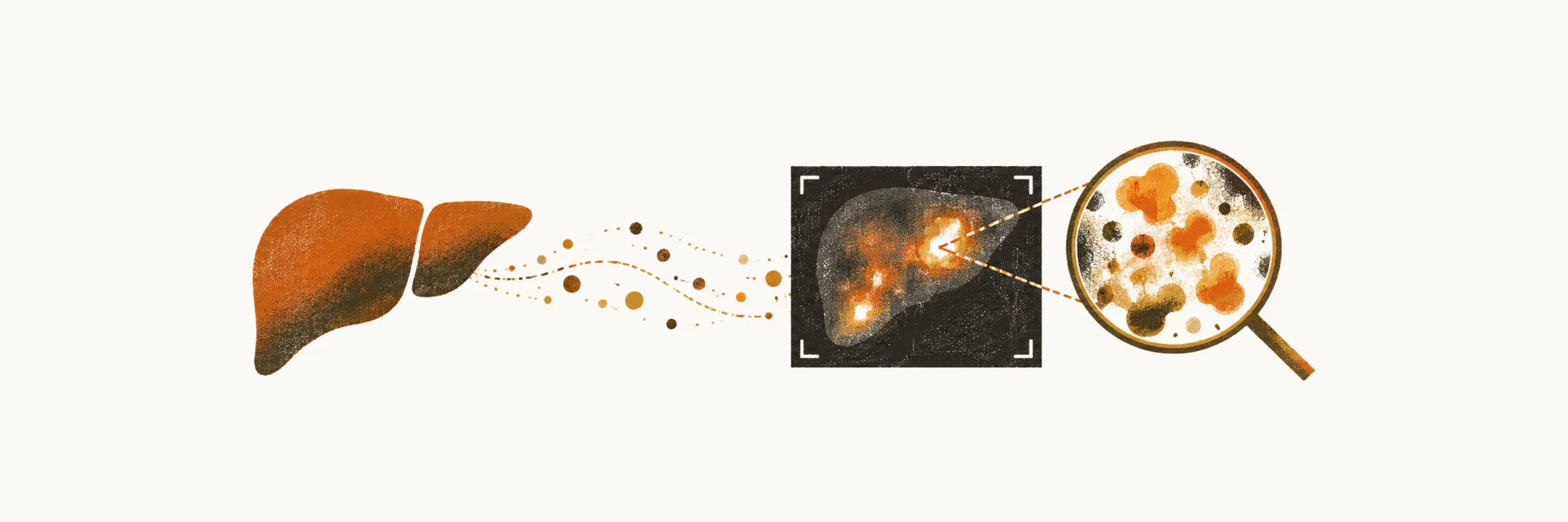
NAFLD has no specific biomarker, so it is usually found by chance and diagnosed by excluding the other conditions that can fill the liver with fat.
Clinical features
Most patients with NAFLD are diagnosed incidentally, because of an increase of liver enzymes on laboratory testing or because of imaging; in most cases the disease is asymptomatic. In symptomatic patients the complaints are:
- vague right upper quadrant (RUQ) pain
- fatigue
- malaise
Stigmata of chronic liver disease, which are ascites, telangiectasia and splenomegaly, can be seen in patients with NASH and cirrhosis.
Diagnosis by exclusion
Because NAFLD does not have any specific biomarker, its diagnosis is based on excluding the other causes of hepatic steatosis, so exclusion of other diseases is one of the main steps in the clinical diagnosis.
One important distinction to make during the diagnosis of NAFLD is its difference from alcohol-associated hepatic steatosis. Based on laboratory findings, in most patients with NAFLD the alanine aminotransferase (ALT) is higher than the aspartate aminotransferase (AST), while in alcoholic patients with alcohol-associated hepatic steatosis the AST level is higher than the ALT level.
Laboratory findings
Studies demonstrated that the mean value of ALT in NAFLD is about 83 IU/ml and of AST about 63 IU/ml. The values of alkaline phosphatase and GGTP (gamma-glutamyl transpeptidase), two enzymes of the bile ducts, may be elevated in these patients, but prothrombin time, albumin level and serum bilirubin, which reflect the liver’s synthetic and excretory function, are mostly in the normal range, except in patients with NAFLD-associated cirrhosis. In about 20–50% of the cases there is an increase of serum ferritin, which can be indicative of more advanced disease.
In about 25% of the cases low-titre positivity for the antinuclear antibody (ANA) can be seen (1:320).
Clinical and laboratory findings in NAFLD are not correlated with the histological findings, so it is possible to have patients who are near-normal on laboratory and clinical findings while cirrhosis is present.
Imaging and biopsy
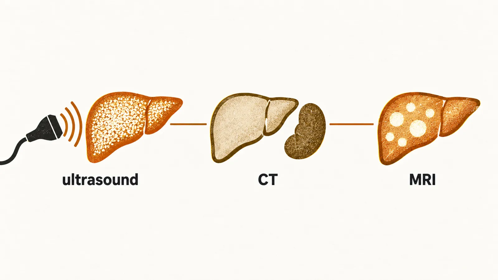
Regarding the imaging studies of patients with fatty liver:
- ultrasound: increased echogenicity, or a bright liver
- CT: the fatty liver appears less dense than the spleen
- MRI: on T1-weighted imaging the fat content can be seen
Diagnosis of NAFLD based on cross-sectional imaging and other laboratory and clinical evidence is easy, but for confirmation of the diagnosis of NASH a liver biopsy has to be performed, and nothing else.
Fibrosis assessment
Because clinical and laboratory findings do not reflect the histology, fibrosis is assessed with dedicated tools. For predicting the fibrosis of the liver there are several modalities:
- FibroScan
- Elasto-MRI
Other than imaging techniques, some biomarkers are indicative of increased fibrosis in the liver, such as CK-18 or NIS4.
The NAFLD fibrosis score has a very good negative predictive value in low scores (the chance that fibrosis is truly absent when the score is low), more than 85%, and also a good positive predictive value in high scores for the presence of fibrosis, which is more than 85%.
The risk of having advanced fibrosis in patients whose AST/ALT ratio approaches 1 is very high, so this value by itself can be used alone for the prediction of advanced fibrosis in patients with NAFLD.