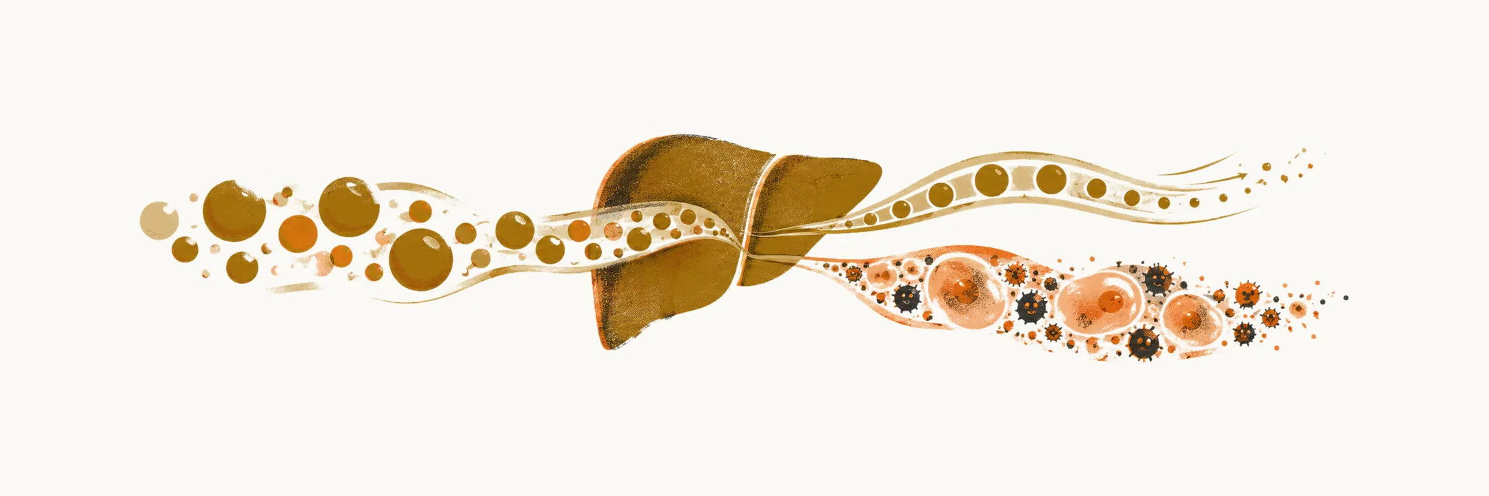
NAFLD covers more than one histological picture, so the terms used to describe it need to be kept distinct. Steatosis means the accumulation of fat inside hepatocytes, the liver’s main cells, and insulin resistance means a reduced response of the body’s tissues to insulin.
The spectrum
NAFLD is a spectrum of disorders characterised by the accumulation of fat inside the hepatocytes, which can be:
- NAFL, or non-alcoholic fatty liver
- NASH, or non-alcoholic steatohepatitis
The defining feature of NAFLD is the accumulation of macrovesicular fat (large fat droplets within the cell) inside more than 5% of the hepatocytes, associated with insulin resistance.
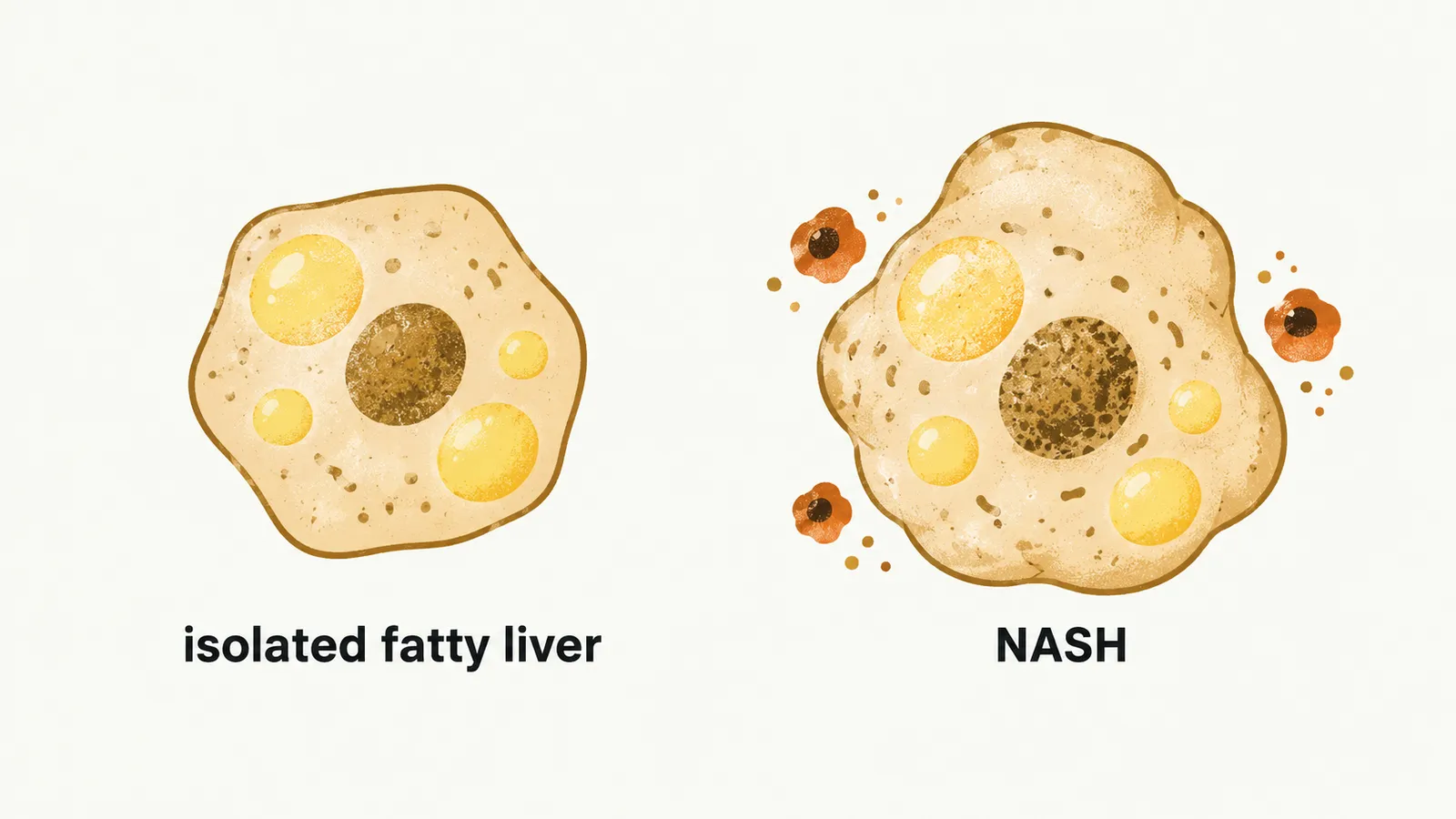
Isolated fatty liver (IFL) is defined as the presence of steatosis in the absence of significant inflammation or fibrosis. The majority of NAFLD is comprised of IFL. NASH is the form in which steatosis is accompanied by inflammation and liver-cell injury.
Nomenclature
The names of these conditions are changing. In 2023 an international consensus replaced NAFLD with metabolic dysfunction-associated steatotic liver disease (MASLD) and NASH with MASH, both to remove the stigma carried by the word “alcoholic” and to build metabolic dysfunction into the definition: MASLD is diagnosed when hepatic steatosis is present together with at least one of five cardiometabolic criteria. The older terms remain in wide use and describe the same spectrum, so both appear in the literature.
Histology of NASH
Under the microscope, NASH is separated from isolated fatty liver by two features. The histological features required to meet the criteria for the diagnosis of NASH are:
- hepatocyte ballooning degeneration
- lobular inflammation with infiltration of mixed inflammatory cells
Other than those two histological features, some other findings can be found in NASH:
- Mallory bodies
- iron deposition
- ductular reaction
- megamitochondria
- vacuolated nuclei in periportal hepatocytes
In patients with NASH, if fibrosis is present it is more commonly located in:
- perisinusoidal regions
- pericellular regions in acinar zone 3
Other causes of hepatic steatosis
A liver that shows steatosis is not necessarily NAFLD. Other conditions can also fill the liver with fat and must be separated from NAFLD. Total parenteral nutrition (TPN), rapid weight loss and starvation each push free fatty acids into the liver, and several drugs do the same. The drugs most often responsible are valproic acid, glucocorticoids, some antiretroviral therapy (ART), amiodarone, tamoxifen and tetracycline.