
Why does the heart suffer most in acromegaly?
The cardiovascular system is where growth hormone (GH) excess has the most significant clinical impact. Cardiovascular disease is the leading cause of death in acromegaly, and more than 60% of the causes of death in these patients are cardiac diseases. Cardiovascular-related comorbidities are present in about 70–90% of patients with acromegaly. They include hypertension, cardiac changes and the other cardiovascular findings of acromegaly.
Acromegaly is usually diagnosed years after it begins, so by the time it is found the heart has already been exposed to excess hormone for years. That exposure acts on the heart muscle itself, which raises a question: how does the hormone reach the heart muscle?
How do GH and IGF-1 act on the heart muscle?
Cardiomyocytes express receptors for GH and IGF-1. Because of these receptors, excess GH and IGF-1 are directly related to cardiac pathology and modification. The myocardium and the endothelium can also make IGF-1 locally in response to GH, which may add to the effect.
Inside the myocyte, the pathophysiology of cardiac hypertrophy is the direct effect of GH and IGF-1 on gene expression. This leads to hypertrophy and increased fibrosis. Sources describe how the two hormones push in the same direction: IGF-1 promotes collagen synthesis by fibroblasts, GH increases cardiac collagen deposition, and IGF-1 also inhibits apoptosis, so myocytes are not lost.
The first phase of cardiac hypertrophy is eccentric hypertrophy of both ventricles. Eccentric hypertrophy means that the ventricular chamber enlarges along with its wall.
These changes do not appear at once; they follow a course.
How does the heart disease progress?
In the early stages of the disease, cardiac modification affects only heart rate and cardiac output. In the later stages the picture becomes congestive heart failure, cardiac hypertrophy and diastolic heart failure. Acromegalic cardiomyopathy has three main stages that link these two ends:
- Stage 1, early, hyperkinetic: heart rate and cardiac output are increased, due to initial cardiac hypertrophy.
- Stage 2, intermediate, hypertrophic: systolic and diastolic dysfunction. Interstitial fibrosis stiffens the wall and impairs relaxation, so filling is impaired first, and the sources describe systolic dysfunction following.
- Stage 3, final, dysfunctional: congestive heart failure and dilated cardiomyopathy.
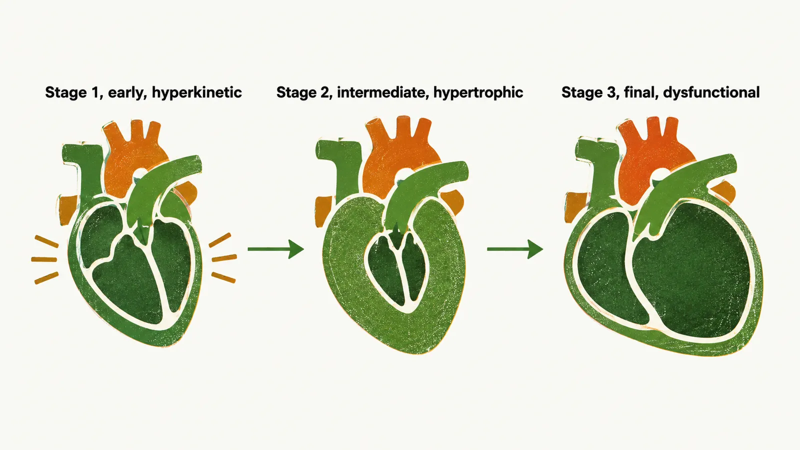
The vessels are changed as well, and pressure rises.
Why does blood pressure rise?
Hypertension is present in 22% of cases, although reported prevalence varies between series. The pathophysiological basis of hypertension in acromegaly is severalfold. The mechanisms are not fully elucidated, but several contribute together:
- Sodium retention and volume expansion. GH acts on the ENaC (epithelial sodium channel) of the renal system, causing sodium retention, and sodium retention leads to volume expansion. Evidence from an animal model and cell experiments suggests that GH switches on this channel in the kidney’s collecting duct, so more sodium is reabsorbed.
- Peripheral resistance is increased. Growth of vessel-wall smooth muscle and greater vascular responsiveness to angiotensin II are proposed contributors.
- Endothelial function is decreased.
- Sleep apnea. Sleep apnea syndrome, common in acromegaly, is a further contributor to hypertension. It arises from the change in airway soft tissue and jaw, and concomitant sleep apnea may exacerbate established hypertension.
Taken together, which cardiovascular problems appear, and which can be undone?
Which findings occur, and which are reversible?
The cardiovascular abnormalities in acromegaly, in order of prevalence, are:
- Hypertension
- Cardiac hypertrophy
- Arrhythmia
- Coronary artery disease
- Systolic heart failure
- Valvular heart disease
Bilateral ventricular hypertrophy and septal hypertrophy are also described, as are valvular diseases such as mitral valve disease and decreased diastolic function. These fit the same picture as the myocyte changes: the walls, the septum and the valve leaflets can all be affected by a disturbed matrix and collagen, with the mitral and aortic valves named in the sources.
Not all of this is permanent. Arrhythmia and cardiomyopathies are reversible after treatment. Valvular heart disease, in contrast, is irreversible after treatment. Wall mass and filling improve with control of GH and IGF-1, and cardiac complications need months of treatment before they improve; the sources give no reason why the valves differ.
If so much of the harm is cardiovascular and part of it can be reversed, how is acromegaly recognised, confirmed and brought under control?