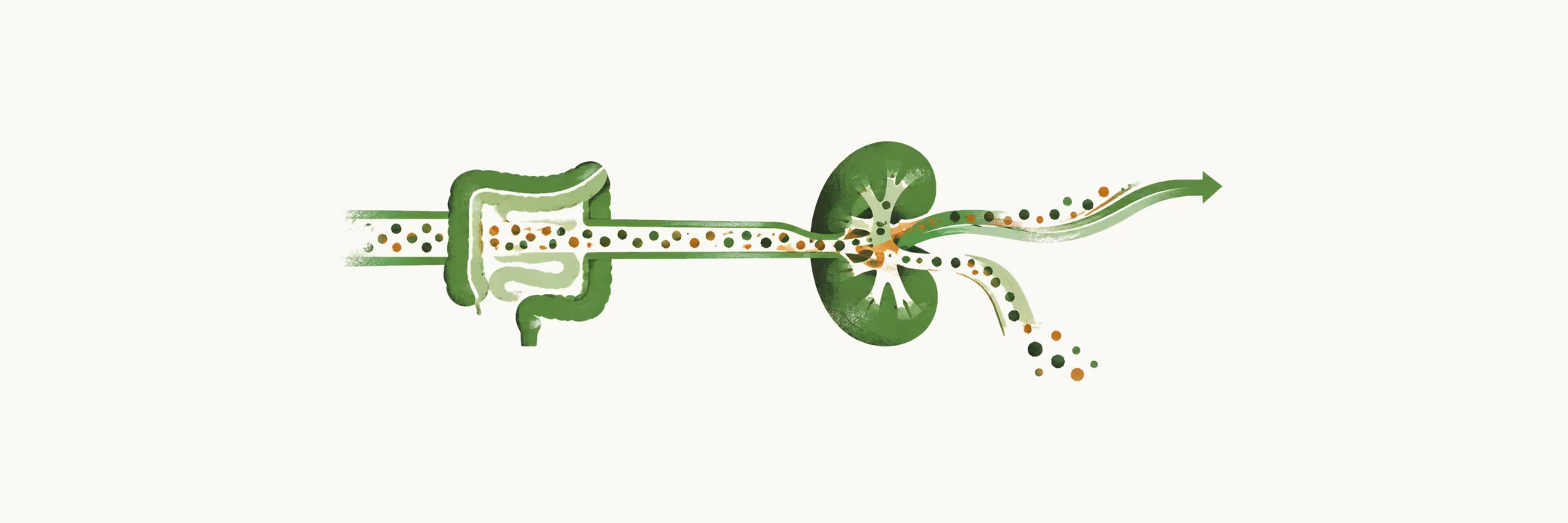
Calcium and phosphate share the same bone and the same organs, but phosphate reaches much further. Phosphate ions are found in bone, in the collagen structure of soft tissues, in cell membranes as phospholipids, inside cells and in the extracellular fluids. More than 80% of the body’s phosphate is in bone, and about 15% is in soft tissues. Phosphate is a constituent of bone mineral, of the phospholipid membrane and of ATP, which is why it is spread through every compartment and not confined to one.
It also does work. Phosphate plays an important role in enzyme activity: kinases use it to activate or deactivate different enzymes. And phosphate forms the energetic bonds of ATP in cells. Because these jobs run through the whole body, a disturbance of phosphate can reach almost any organ system.
Why is phosphate controlled less tightly than calcium?
If phosphate matters this much, why does its level wander with the meal? Phosphate homeostasis is not controlled as tightly as calcium concentration in the body, and phosphate concentration shows quite variable perturbation during the day, based on diet. Part of the reason is that diet supplies phosphate far beyond the daily need; the amount in different diets is much higher than needed, and even inadequate diets contain a lot. The other part is that one of the intestinal absorption routes cannot be turned down. Because absorption is not matched to need, the level follows intake.
What does phosphate do in the blood?
Besides its structural and energy roles, phosphate has one regulating-like job in plasma: buffering. At blood pH 7.4, the phosphorus of the extracellular fluid is in two forms, NaH2PO4 and Na2HPO4, and Na2HPO4 is four times more abundant than the monosodium form. The two forms interconvert at a pH close to that of blood. A buffer pair works best when the pH lies near the point where its two forms interconvert, so the pair can take up or release hydrogen ion without a large change in pH. That is how the pair buffers the extracellular fluid, and in extracellular fluids phosphate does act as a buffer.
How well does it do the job? Buffering depends on concentration as well as on a good match with the pH. Because of its low concentration, phosphate is not considered the main buffer system of the blood.
Why does the gut not set the balance?
Given that so much phosphate is available, the next question is where the balance is set. Most phosphate ion, about two thirds, is absorbed by the intestine, and it crosses by two molecular mechanisms.
The first is passive absorption. A passive route follows the concentration gradient and has no regulatory switch, and this mechanism is almost always not modulated. Because passive transporters are not downregulated, phosphate keeps entering even when the body is overloaded, and some patients have persistent hyperphosphatemia. The second is active, vitamin-D dependent absorption; as with calcium, calcitriol drives this route.
Put the pieces together. Intake exceeds need, and the passive route is not turned down, so the intestine does not set the balance. The balancing mechanism that keeps phosphate in the body rests on the kidney, because renal excretion is the adjustable step that remains.
How does the kidney set the balance?
In the physiological state, almost all absorbed phosphate is excreted in the urine, so the kidney sets the level by deciding how much to keep. In hyperphosphatemia, the phosphate transporters of the renal tubules are down-regulated; fewer transporters mean less reabsorption and more phosphate in the urine, which is the kidney’s response to surplus.
As with calcium, the kidney filters a large load and takes most of it back. More than 90% of free plasma phosphate is filtered into the urine, and more than 80% of the filtered phosphate is reabsorbed in the proximal convoluted tubule (PCT). The PCT is the main site of reabsorption, so a small change in reabsorption there changes the amount excreted by a large factor. Renal reabsorption is therefore the step that sets the phosphate balance.
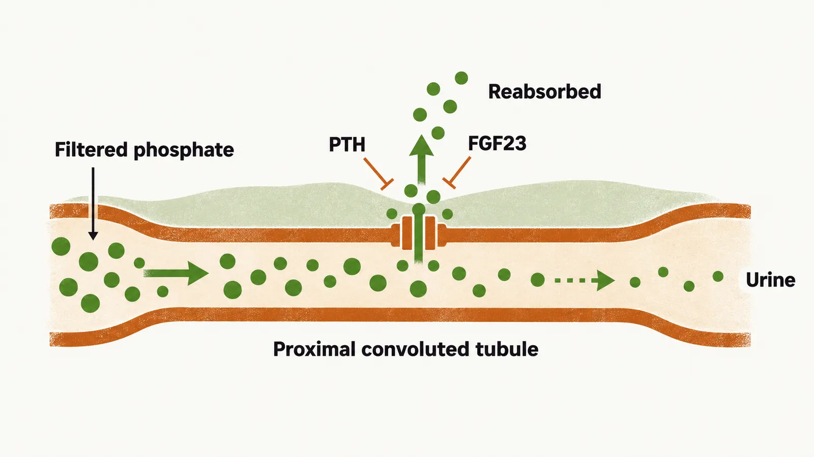
What controls that step? Renal phosphate reabsorption is under the control of:
- PTH and FGF23, the hormones that control renal phosphate reabsorption;
- dietary phosphorus intake;
- non-hormonal factors: extracellular volume and acid-base status.
Expansion of plasma volume, for example, increases phosphate excretion.
What does phosphate do in the urine?
The phosphate that is not reabsorbed stays in the tubular fluid and has a physiological effect in the urine itself. Phosphate is accumulated in the urine as it passes toward the distal tubules, where a proton ion is exchanged with sodium. There, Na2HPO4 is converted to NaH2PO4. The proton secreted by the tubular cell binds a base other than bicarbonate, here phosphate, so acid leaves as phosphate-bound proton and not as free acid. This conversion permits urine to accumulate as much proton ion as needed without any change in urine pH.
So phosphate is not the main buffer in blood but a useful one in the urine. If renal reabsorption sets the balance, what do PTH and FGF23 do to it, and how does the Parathyroid Hormone (PTH) system sense that calcium has fallen?