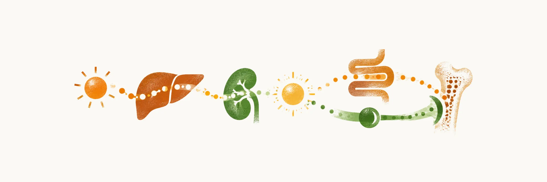
Vitamin D is more a hormone than simply a vitamin and co-factor. The form that arrives from skin or diet does nothing by itself. The body activates it in two steps, one in the liver and one in the kidney, and the kidney step is where the amount of active hormone is set. The active hormone, calcitriol, acts on gut, kidney and bone far from where it is made. How calcitriol itself is made is the question the FGF23 story leaves open.
Where does vitamin D come from, and why is it called a hormone?
The term vitamin D covers two fat-soluble compounds: vitamin D3, cholecalciferol, and vitamin D2, ergocalciferol. The two names follow two origins: D3 comes from 7-dehydrocholesterol in the skin, and D2 from a plant and yeast sterol.
D2 and D3 differ less in what their active metabolites do than in how the body handles them. The metabolic steps of D2 are similar to those of D3, and their active metabolites have comparable biological activity at the vitamin D receptor. D3 binds the carrier protein, vitamin D binding globulin, more tightly than D2, and it has a longer half-life. Because of this tighter binding and longer half-life, D3 raises the circulating 25-hydroxy level more effectively than D2.
Vitamin D is also unusual among vitamins. The skin produces its precursor, 7-dehydrocholesterol, and in response to skin exposure to light this is converted to cholecalciferol. So the compound can be made, rather than only absorbed.
What is not made in the skin has to be absorbed, and because vitamin D is fat-soluble, absorption happens in the small intestine together with dietary fat. Bile is needed for this absorption and also for the excretion of vitamin D. That explains why intestinal inflammation can reduce vitamin D absorption, and why hepatic and biliary disorders lower it: both interfere with taking up a fat-soluble vitamin.
Vitamin D from skin or gut is biologically inert. How does it become the active hormone?
How is vitamin D activated?
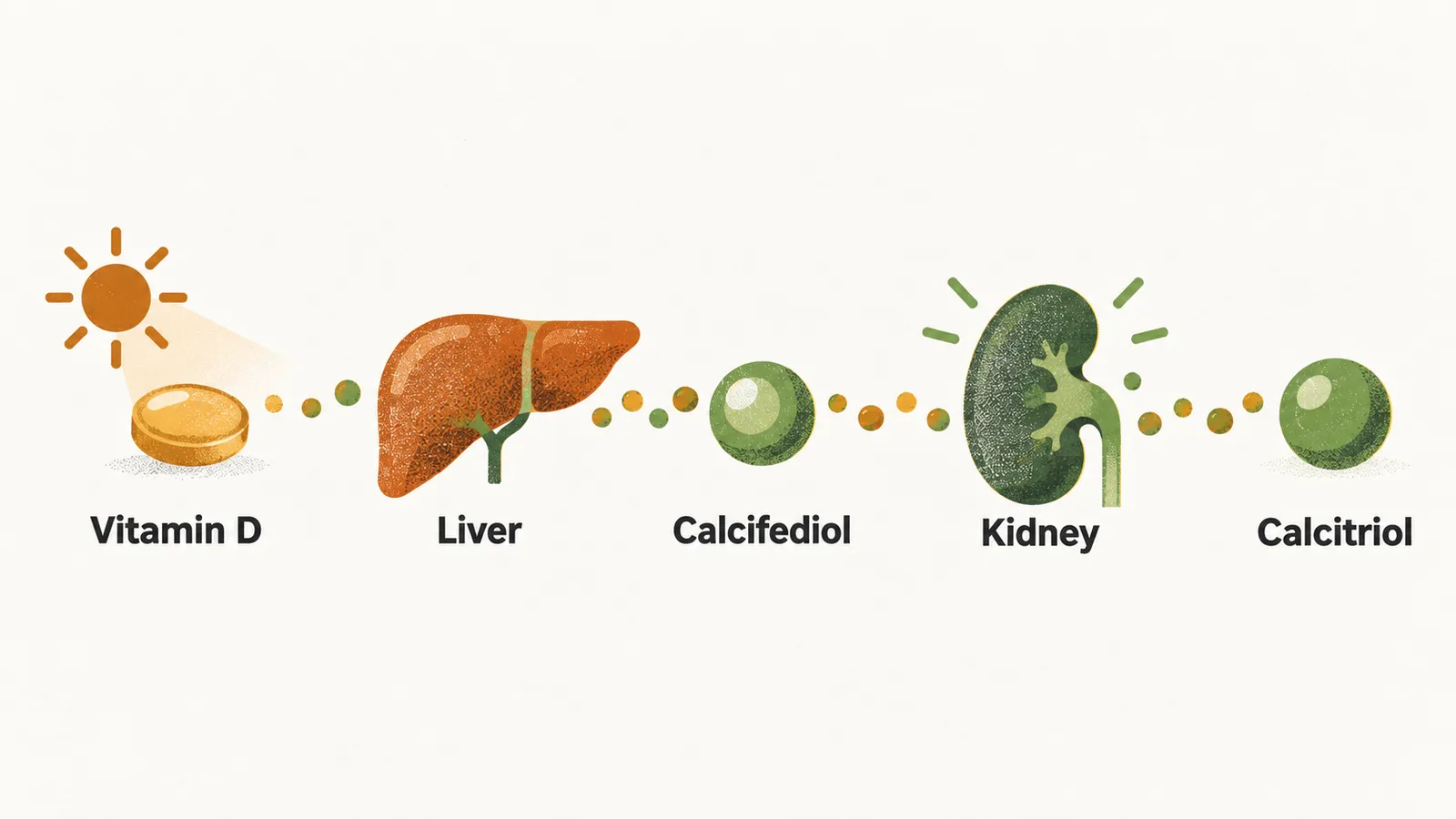
Vitamin D requires activation by successive hydroxylation steps. The first happens in the liver: 25-hydroxylation of cholecalciferol forms 25-hydroxycholecalciferol, or calcifediol. This step is not tightly regulated and is limited mainly by how much vitamin D arrives, so the circulating level of calcifediol follows what the skin makes plus what the diet supplies.
Calcifediol is the major circulating form of vitamin D. It is carried mainly on vitamin D binding globulin, which is how a fat-soluble molecule travels in blood. Its half-life is about 19 days, long enough to make it a stable store.
The second step happens in the kidney. Calcifediol bound to its carrier is filtered at the glomerulus and taken up by cells of the proximal convoluted tubule, and there 1α-hydroxylation of 25-hydroxycholecalciferol forms 1α,25-(OH)2-cholecalciferol, or calcitriol. Calcitriol is the most potent and biologically active form of vitamin D; it is the form that binds the vitamin D receptor. Its half-life is only about 4 to 6 hours. With a short half-life and tightly regulated production, the body can adjust the amount of active hormone quickly, and a calcitriol level is a poor marker of the vitamin D stock, which is held as calcifediol.
The liver step runs on whatever supply arrives, so the kidney step is the one the body can control. What does it respond to?
What raises or lowers calcitriol production?
Calcitriol production is highly regulated by dietary calcium intake, by the calcium level, by the phosphate level, by the FGF23 level and by the calcitriol level itself.
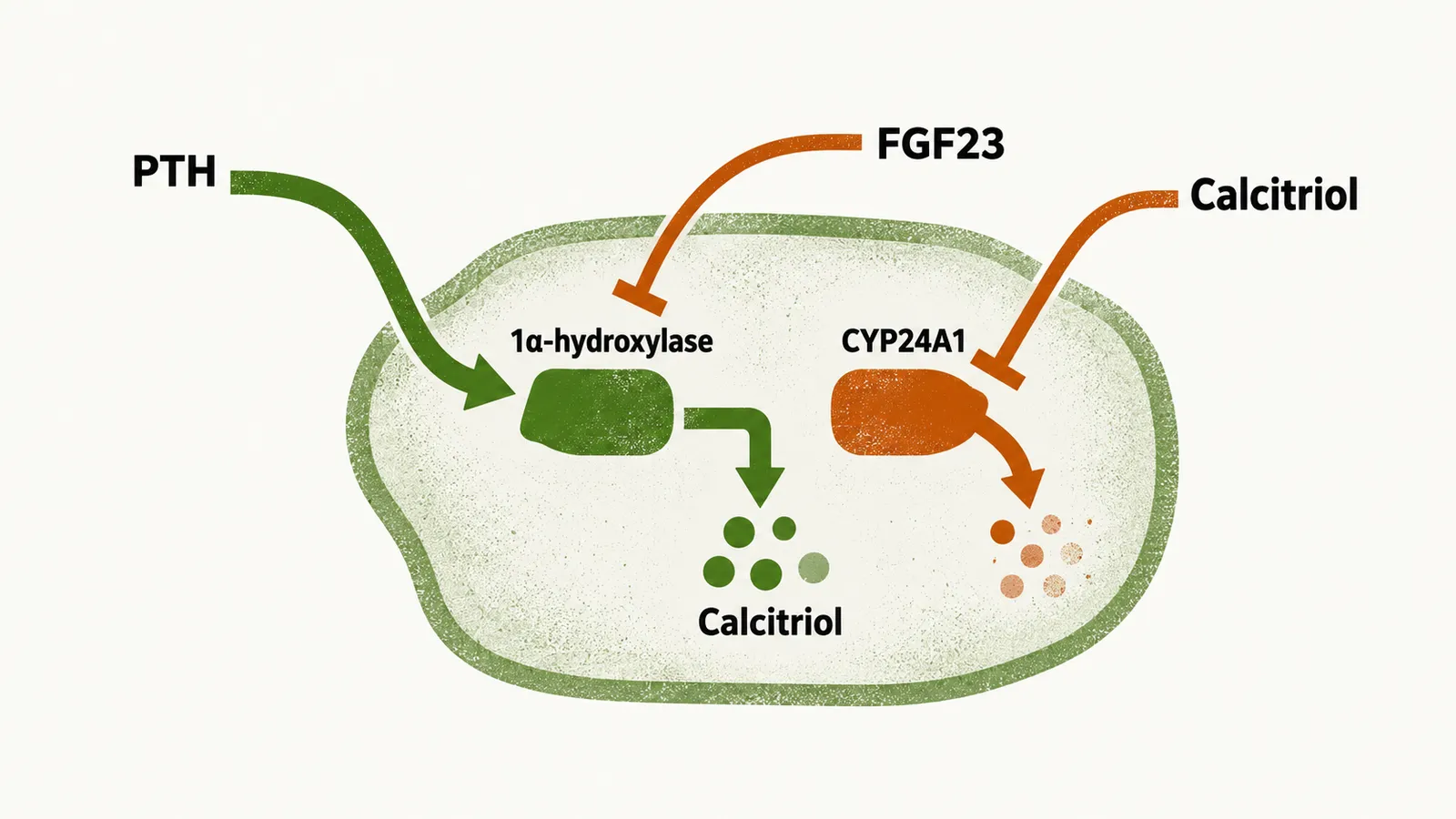
The kidney controls production by balancing two opposed enzymes. 1α-hydroxylase switches vitamin D on. The CYP24A1 enzyme switches it off: 24-hydroxylation of calcifediol forms 24,25-(OH)2-cholecalciferol, an inactive metabolite, which cannot bind the vitamin D receptor. This switch-off step acts on calcitriol and on calcifediol alike. Each input works by turning one enzyme up or the other down.
- Calcium and PTH. PTH switches on 1α-hydroxylase in the proximal tubule, so low calcium raises calcitriol through PTH. A high calcium level counteracts the PTH effect. PTH also down-regulates CYP24A1, so it raises calcitriol by both routes: more activation and less breakdown.
- Phosphate. Low phosphate raises calcitriol production independently of PTH; the mechanism of this effect is not known.
- FGF23. FGF23 restrains calcitriol formation, and calcitriol in turn drives FGF23, a loop. FGF23 lowers calcitriol by suppressing the activating enzyme and upregulating CYP24A1.
- Calcitriol itself. Calcitriol lowers its own production in the same way, a negative feedback that keeps it from running away. It upregulates CYP24A1, which shifts the balance toward breaking calcitriol down.
The regulation can be bypassed. In some diseases 1α-hydroxylase is over-expressed by macrophages, for example in sarcoidosis, or by lymphocytes, for example in lymphoma. These cells make calcitriol without the kidney’s feedback, so production runs unregulated, and this over-expression leads to hypercalcemia.
With calcitriol made and controlled, what does it do?
What does calcitriol do?
Calcitriol’s physiological effect is increased absorption and retention of calcium and phosphate in kidney and intestine: the gut takes up more, and the kidney loses less. Part of intestinal phosphate uptake is vitamin D dependent, and the proximal tubule reabsorbs most filtered phosphate.
- Intestine. Calcitriol increases intestinal absorption by augmenting active, vitamin-D-dependent calcium absorption in the proximal duodenum, raising the gut’s own calcium transport machinery.
- Kidney. Calcitriol decreases renal excretion. It acts on the distal tubule, the regulated step of calcium reabsorption. For phosphate in the proximal tubule it is thought to act similarly, but the mechanism there requires further elucidation.
Bone is the calcium store, and calcitriol also enhances bone mobilization. Together with PTH, it drives the formation of mature osteoclasts, with the osteoblast signalling the osteoclast, so that bone releases calcium and phosphate into the blood when the diet cannot supply enough. Calcitriol is a bone-mobilizing hormone, not a bone-forming hormone. By raising calcium and phosphate in the blood it can support mineralisation indirectly, but it is not itself a bone-forming hormone.
The osteoblast carries a calcitriol receptor that promotes the formation of some mediators. Osteocalcin promotes bone formation, whereas IL-1 promotes bone resorption. IL-1 and osteocalcin have opposite effects on bone remodeling, so calcitriol is better described as bone-mobilizing than bone-forming.
Everything so far pushes calcium up. What pulls it down?
What brings calcium down?
Calcitonin is the counterpart that lowers calcium: a hypocalcemic hormone. It is released when plasma calcium concentration is high, so it is the high-calcium arm that balances PTH. The parafollicular cells of the thyroid gland release it in response to high calcium.
Its target is the osteoclast. Calcitonin is the most potent peptide inhibitor of osteoclast-mediated bone resorption: its receptor is on the osteoclast, and calcitonin makes the osteoclast contract, so it moves less and resorbs less bone. It also hinders osteoclast precursors from maturing.
Normal calcitonin is less than 25 pg/mL in men and less than 20 pg/mL in women, so the two sexes have different normal ranges. Abnormally high calcitonin is seen in medullary thyroid carcinoma (MTC).
The hormones so far act on bone, kidney and intestine to raise or lower calcium in response to the plasma level. But calcium can also rise when a tissue makes a peptide that acts on the PTH receptor, whatever the state of the parathyroid glands. What is that peptide, and how does it raise calcium without the parathyroid?