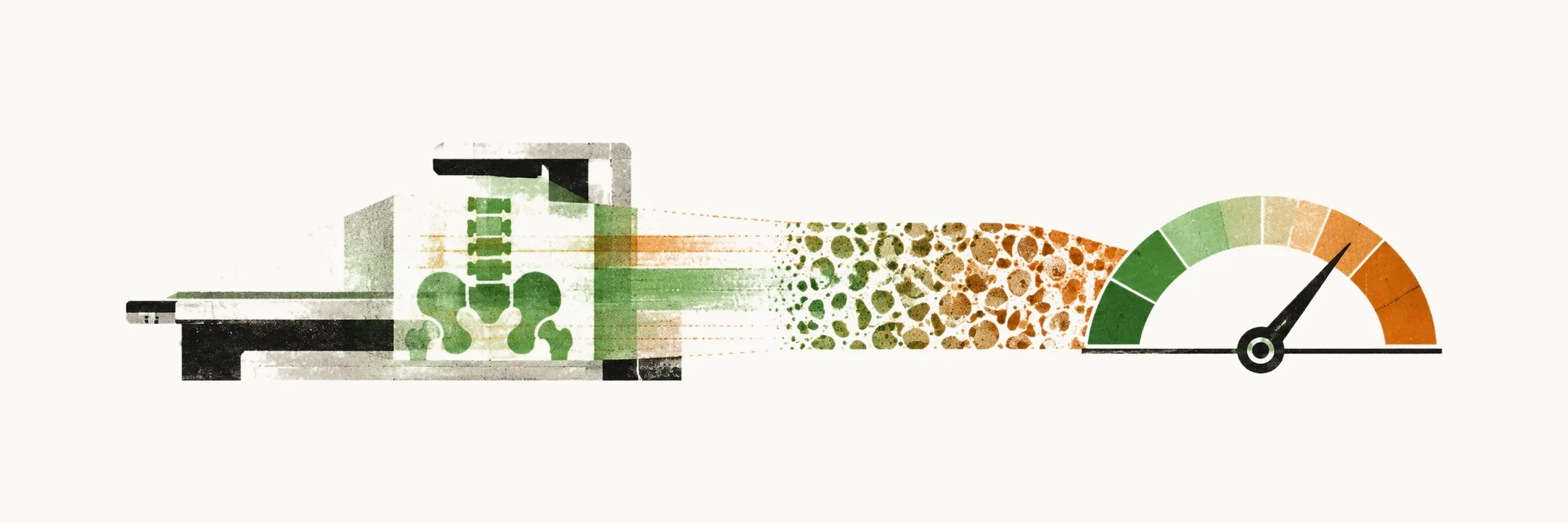
Diagnosing osteoporosis has two parts. One confirms that bone mineral density is low. The other recognises a fracture that may already have occurred.
The disease is silent. Bone loss gives no symptoms of its own, as the pathophysiology note explained. So the diagnosis rests on measurement and on imaging of symptomless vertebral fractures, not on how the patient feels.
What does the examination look for?
The examination of a patient with suspected osteoporosis comes before any scan, and it has two purposes. The first is to find signs of a secondary cause. The second is to detect a vertebral fracture or a high risk of falling.
Signs of another cause of bone loss
Some signs point to a disease that may be behind the bone loss.
- An enlarged thyroid gland suggests hyperthyroidism. Overt hyperthyroidism is an established cause of high bone turnover with accelerated bone loss, because thyroid hormone speeds up bone turnover.
- Atrophic testicles suggest hypogonadism. Estradiol is the key sex steroid for the skeleton in both sexes, as the pathophysiology note explained.
- Blue sclera with yellow-brown teeth and hearing loss suggest osteogenesis imperfecta.
Signs of falling and high fracture risk
A body mass index below 20, or weight loss of more than 5%, suggests a high risk of fracture.
Abnormal posture and gait, reduced muscle strength, postural hypotension and impaired consciousness suggest a higher risk of falling. This matters because falls and trips magnify force on a small area of the proximal femur, as the epidemiology note explained.
Signs of a vertebral fracture
A vertebral fracture is easy to miss, so the examination looks for its signs. Localised tenderness over a spinous process, kyphosis, or a reduced distance between the lower ribs and the pelvic brim suggests a vertebral fracture. Collapsed vertebrae produce kyphosis, as the clinical manifestation note explained.
Who should have a density scan?
The examination shows the signs. The next question is who should have a scan at all. The US BHOF recommends BMD testing in these groups:
- Women aged 65 years or more and men aged 70 years or more: consider BMD testing.
- Postmenopausal women and men aged 50-69 years: consider BMD testing based on risk profile. The steep loss of bone in the years after the menopause comes from estrogen loss, as the pathophysiology note explained.
- Postmenopausal women and men aged 50 years or more with a history of fracture in adult age: consider BMD testing. A fracture after the age of 50 is a sentinel warning, as the clinical manifestation note explained.
- Adults with conditions (for example rheumatoid arthritis, organ transplant) or medications (for example glucocorticoids, aromatase inhibitors, androgen deprivation therapy) associated with low bone mass or bone loss: consider BMD testing. Glucocorticoid-induced bone loss, with its time course and routes, was taught in the pathophysiology note.
How is bone density measured?
Once a patient is chosen, the scan measures the bone at fixed sites. BMD is measured by DXA, the scan described in the fracture-risk note.
DXA should sample the lumbar spine (L1-L4), the femoral neck, the total hip and the 33% radius. The forearm is the fallback, because forearm DXA may be obtained when the lumbar spine or hip cannot be measured.
What does the T-score mean?
The scan gives a density, and the T-score says how far that density is from the young adult average. The T-score is the number of standard deviations by which the patient’s BMD, measured by DXA, differs from the mean of the young adult population. Peak bone mass is reached in young adult life and bone loss comes after it, as the pathophysiology note explained.
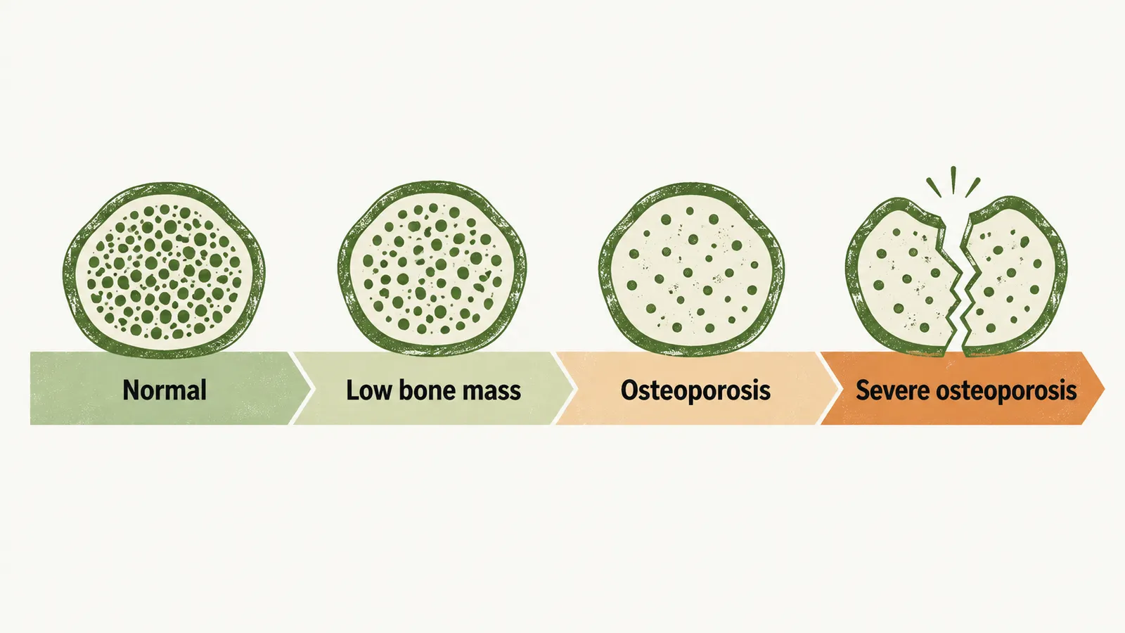
The WHO defines osteoporosis from the T-score:
- Normal: T-score of -1.0 or greater.
- Low bone mass (osteopenia): T-score between -1.0 and -2.5.
- Osteoporosis: T-score of -2.5 and below.
- Severe osteoporosis: T-score of -2.5 and below plus a fragility fracture.
Can a fracture diagnose osteoporosis without a scan?
The T-score needs a scan, but a fracture can also count as evidence. A fragility fracture is a fracture that occurs with little force. A weakened bone breaks under forces a healthy bone survives, as the epidemiology note explained.
So with a fragility fracture, osteoporosis may be diagnosed even without measuring BMD, provided the fracture is not explained by another cause such as multiple myeloma. The condition matters because a tumour can weaken bone too. In myeloma-related bone disease the bone-remodelling balance is upset by osteoclast activation and osteoblast suppression. A fracture is therefore not proof of osteoporosis until another cause is excluded.
Why is the spine imaged?
Most vertebral fractures are silent, as the clinical manifestation note explained, so a fracture that causes no pain is not reported and has to be looked for with imaging.
Vertebral fracture assessment (VFA) uses the same machine as DXA, with lateral imaging, to find vertebral fractures. The DXA machine can image the spine from the side during the regular BMD measurement, so the same scan can look at the vertebrae.
How is vertebral height used?
A loss of height is the first clue, and the image then gives the number and the severity. A collapsed vertebral body is shorter than it was, and the spine loses that height. Two measurements of height follow from this:
- A loss of more than 4 cm from the tallest remembered height suggests an old vertebral fracture.
- A loss of more than 2 cm from a previous height measurement suggests a new vertebral fracture.
VFA is semi-quantitative: the height of each vertebral body is measured. Each body is measured at its front, middle and back, so a wedge or collapse shows as a difference between them. Severity is graded by how much vertebral height is lost, so the reduction in height gives the number and severity of the fractures.
When is lateral spine imaging used?
Lateral imaging is used in some people and not in others, depending on the T-score and other features. VFA is indicated when the T-score is below -1.0 and at least one of the following is also present:
- Age: women aged 70 years or more, or men aged 80 years or more.
- Historical height loss of more than 4 cm (1.5 inches).
- A self-reported but undocumented prior vertebral fracture.
- Glucocorticoid therapy equivalent to 5 mg of prednisone or more per day for 3 months or more.
How do density and risk combine?
The T-score is one input, and the decision also depends on the other risk features. Density is not the whole of bone strength, as the fracture-risk note explained. FRAX is the tool that combines BMD with clinical risk factors.
The BHOF treats the diagnosis as definite in a postmenopausal woman or a man older than 50 years in two situations:
- A DXA T-score of -2.5 or lower.
- A DXA T-score between -1.0 and -2.5, and a FRAX 10-year probability of major osteoporotic fracture above 20% or of hip fracture above 3%.
What if the findings point to another cause?
A fracture or a sign can point away from age, towards a disease, deficiency or drug. Findings such as an enlarged thyroid gland or atrophic testicles raise the question of which other diseases, deficiencies or drugs may be behind the bone loss. A fracture that another cause such as myeloma could explain raises the same question. So which other diseases, deficiencies or drugs may be behind this bone loss, and how are they found?