What does an excess of glucocorticoid look like, and why is it so variable?
Cushing’s syndrome is defined by chronic excess of glucocorticoids. Glucocorticoid receptors are present in most cells of the body, so the excess reaches almost every tissue and produces a wide, variable set of manifestations. The variability is itself part of the clinical problem. Most individual findings are also common in the general population, so the syndrome is recognised by the pattern, not by any single symptom.
How often is each finding seen?
Reported prevalence differs by finding; weight gain appears as a presenting feature.
| Finding | Prevalence |
|---|---|
| Weight gain (presenting feature) | 81-97% |
| Round face | 88-92% |
| Hypertension | 68-90% |
| Skin thinning | 84% |
| Hirsutism | 56-81% |
| Dysmenorrhea | 35-84% |
| Purple wide striae | 35-84% |
| Reduced libido | 33-100%, higher in men |
| Muscle weakness/tiredness | 46-67% |
| Diabetes/impaired glucose tolerance | 43-50% |
| Edema | 48-50% |
| Mental health disorders | 26-62% |
| Easy bruising | 21-62% |
| Fractures | 21-56% |
| Acne | 19-64% |
| Female balding | 13-51% |
| Recurrent infections | 14-25% |
Which of these numbers point to the syndrome?
Which findings are non-specific and which are specific?
The majority of signs and symptoms are non-specific. Hirsutism with frontal balding, diastolic hypertension and depression are examples, and all of them also occur without Cushing’s syndrome. Because each is so common alone, the syndrome should be suspected when several are present simultaneously at an unexpected age, for instance osteoporosis in a young adult.
Specific features are more useful, and they reflect loss of tissue: fragility and thinning of the skin, easy bruising with broad lesions larger than 1 cm, and proximal myopathy. Because the specific findings are the ones that reflect tissue breakdown, what is being broken down?
Why does cortisol break down muscle, skin and bone?
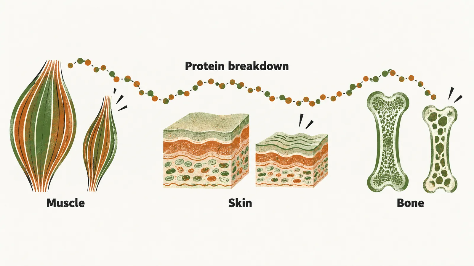
The most prominent features relate to increased lipolysis and protein catabolism. Cortisol drives the liver to make glucose from amino acids, and protein is broken down to supply those amino acids. This fits the impaired glucose tolerance seen in 43-50% of patients, as a link rather than a proven sole cause.
The tissues that depend on protein pay for it:
- Muscle. Muscle breakdown produces proximal myopathy, and also the weakness and tiredness patients report.
- Skin. The same process thins the dermis. That explains the skin fragility, the purple striae and the easy bruising.
- Bone. Bone matrix is protein, so protein catabolism contributes to fractures.
Fat is redistributed to the trunk, face and dorsocervical region. So weight gain comes with a round face and truncal obesity, and it is not a uniform accumulation of fat.
Not every feature follows from breakdown.
Where do hirsutism, acne and balding come from?
Hirsutism, acne and female balding follow a different route. In ACTH-dependent disease and in ectopic carcinoma, hyperandrogenism is common, because high ACTH also drives adrenal androgen production alongside cortisol. In ACTH-independent disease, where ACTH is not driving the adrenal, it is mostly only the glucocorticoid that is in excess.
Another group of features comes from a different receptor altogether.
Why can cortisol act like aldosterone?
The mineralocorticoid receptor is normally aldosterone’s, and through it aldosterone retains sodium and loses potassium. Cortisol and aldosterone both activate this receptor. The kidney protects the receptor with the enzyme 11β-hydroxysteroid dehydrogenase type 2 (11β-HSD2), which converts cortisol to inactive cortisone before it can act.
When cortisol production is high enough, the enzyme can no longer keep up. Cortisol escapes inactivation and exerts mineralocorticoid effects. The result is hypokalemia, together with diastolic hypertension and edema; the edema is consistent with sodium retention. In ectopic ACTH, serum potassium is less than 3.3 mmol/l in more than 70% of cases. The mineralocorticoid effect appears when urine cortisol is more than 4100 nmol/day.
Cortisol also reaches the brain.
What does high cortisol do to the hypothalamus, pituitary and blood count?
High cortisol also acts centrally on the hypothalamus and pituitary. It suppresses the gonadotropins. Experimental work indicates that glucocorticoids suppress gonadotropin release at the pituitary and GnRH release at the hypothalamus. The outcome is hypogonadotropic hypogonadism, with amenorrhea, hypogonadism and an irregular menstrual cycle. In the same way, cortisol suppresses the hypothalamic-pituitary-thyroid (HPT) axis, giving a low TSH.
Cortisol also changes the blood count. Leukocytosis can occur in Cushing’s syndrome, because neutrophils are demarginated, that is released from the vessel wall where they had been resting, into the circulating pool.
Do any signs point to the cause?
Which signs hint at ectopic ACTH?
Mostly, signs and symptoms do not separate the different etiological causes. A few hints point toward ectopic ACTH: hyperpigmentation of the knuckles, and rapid progression of edema, hypokalemia and hypertension.
Hyperpigmentation is a consequence of POMC, the precursor from which ACTH is cleaved. Alongside ACTH, the tumour processes peptides that stimulate skin melanocytes, so a tumour producing large amounts of POMC darkens the skin.
Ectopic ACTH-dependent Cushing’s syndrome mostly presents with severe and rapidly progressive signs and symptoms, for example anorexia, myopathy, glucose intolerance and hypokalemic alkalosis. Rapid deterioration, hypokalemic alkalosis, knuckle hyperpigmentation and clinical signs of hypercortisolemia together should trigger high suspicion of small cell lung carcinoma with ACTH secretion or high-grade bronchial carcinoma.
Sodium retention, hypertension and fluid-laden tissues raise a further question: what does all this do to the heart and vessels?