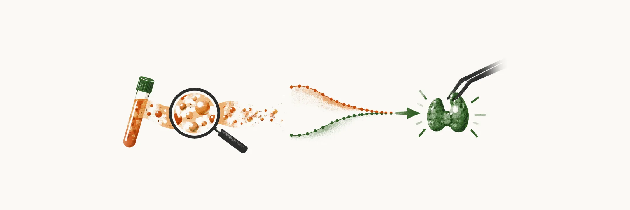
Diagnosing PHPT means showing hypercalcemia together with a high, or inappropriately normal, PTH level, and then excluding other causes of that picture. The diagnosis therefore rests on two grounds: high calcium with high PTH, or high calcium with an inappropriately normal PTH.
Diagnosis here is a comparison, not a single number. Calcium is the reference: a healthy gland should answer a high calcium with a lower PTH. When PTH stays high, or sits inside the range when it should be lower, the gland is the likely source. The question then becomes which other cause could produce the same pair of values. Each test in the workup answers one part of that question, and the pattern of calcium, PTH, phosphate, urine calcium and bone changes points to one cause or another.
What does a high PTH with a high calcium show?
This is the plain ground. The serum calcium is high and the PTH is high too, usually within 2 times above ULN.
Why is a normal PTH a clue when calcium is high?
The second ground needs a precise meaning of “inappropriately normal or elevated”. A healthy gland turns PTH down when calcium is high, and PHPT is a shifted set point, as described in Epidemiology and pathogenesis of primary hyperparathyroidism.
So physiologically, in hypercalcemia PTH is suppressed to the lower limits of normality or even lower. That is why, in hypercalcemia, a PTH within the normal range of 10-65 pg/ml is not normal. It is inappropriately normal, and it supports the diagnosis just as a high value does.
Why is the PTH range hard to read?
A PTH inside the range still needs context, because the range of 10-65 pg/ml is quite wide. Three things widen it:
- Age. The wide range reflects physiological change, because the PTH level increases within the normal range with age.
- Vitamin D level. The normal range is extended in vitamin D deficient populations.
- Renal dysfunction. PTH levels are higher because of abnormal clearance and degradation of PTH.
What else changes besides calcium and PTH?
Calcium and PTH are not the only changes; phosphate and urine calcium move too.
Phosphate is low, but within the normal range. This follows from the action of PTH on the renal tubule: PTH blocks phosphate reabsorption, so the kidney loses more phosphate and blood phosphate falls. Frank hypophosphatemia happens in only 25% of cases.
Hypercalciuria is present in 40% of cases. The kidney filters more calcium than it reclaims, so urine calcium runs high, as the pathogenesis note explains.
How does vitamin D change?
The same PTH that raises calcium also changes the vitamin D values. In 30% of cases, 25-hydroxy vitamin D is in the low-normal range, and 1,25-dihydroxy vitamin D is in the upper-normal range or increased.
Both are caused by the effect of PTH on 1α-hydroxylase of the renal tubules. This kidney enzyme converts 25-hydroxy vitamin D to calcitriol (1,25-dihydroxy vitamin D). When PTH drives the enzyme, more of the substrate is turned into product, so calcitriol runs high-normal or high. The low-normal substrate is probably a consequence of that use. How PTH raises calcitriol is described in Clinical presentation of primary hyperparathyroidism.
What do the measured values look like?
These are the values the changes above produce. The table gives the mean ± SEM in a group of PHPT patients, set beside the normal ranges.
| Test | PHPT patients | Normal range |
|---|---|---|
| Serum calcium | 10.7 ± 0.1 mg/dL | 8.2-10.2 mg/dL |
| Serum phosphorus | 2.8 ± 0.1 mg/dL | 2.5-4.5 mg/dL |
| Total alkaline phosphatase | 114 ± 5 IU/L | <100 IU/L |
| Serum magnesium | 2.0 ± 0.1 mg/dL | 1.8-2.4 mg/dL |
| PTH (IRMA) | 119 ± 7 pg/mL | 10-65 pg/mL |
| 25(OH)D | 19 ± 1 ng/mL | 30-80 ng/mL |
| 1,25(OH)2D | 54 ± 2 pg/mL | 15-60 pg/mL |
| Urinary calcium | 240 ± 11 mg/g creatinine | none listed |
| Urine DPD | 17.6 ± 1.3 nmol/mmol creatinine | <14.6 nmol/mmol creatinine |
| Urine PYD | 46.8 ± 2.7 nmol/mmol creatinine | <51.8 nmol/mmol creatinine |
Alkaline phosphatase, DPD and PYD are products of bone-forming and bone-removing cells, so they show the effect on bone, which Bone disease in primary hyperparathyroidism explains.
Which other causes give the same pattern?
A high calcium with a high PTH has more than one cause. Once hypercalcemia with high or inappropriately normal PTH is found, other forms of hyperparathyroidism and other causes of hypercalcemia have to be separated from PHPT. Each cause has its own biochemical pattern.
In PHPT the serum calcium is elevated, PTH is elevated or inappropriately normal, and urine calcium is high or high normal. That is the pattern the other causes are measured against.
How does hypercalcemia from cancer differ?
The first cause to separate is the one in which the gland’s hormone is quiet. One of the most important steps in the differential diagnosis of PHPT is hypercalcemia of malignancy. In hypercalcemia of malignancy and in non-PTH mediated hypercalcemia, serum calcium is elevated, PTH is suppressed, and urine calcium is typically high.
Usually these patients are symptomatic, with severe hypercalcemia and a decreased serum PTH. Decreased PTH should accompany hypercalcemia unless PTH is itself the cause, so a suppressed PTH points away from PHPT.
In some malignancy-related hypercalcemia there is increased production of PTHrP (PTH-related protein). PTHrP is a related hormone that cross-reacts with the PTHR-1 receptor, the receptor PTH acts on. It raises bone resorption and renal calcium reabsorption and so leads to hypercalcemia, while the native PTH stays suppressed.
More than 90% of hypercalcemia cases are related to either PHPT or malignancy.
Do drugs and other causes need checking?
Some causes are drugs, which can be checked by withdrawing them. The other possible causes for the differential diagnosis are:
- vitamin D intoxication
- granulomatous disease
- FHH
- lithium use
- thiazide use
Lithium shifts the same set point that PHPT shifts. If withdrawing lithium or thiazide corrects the hypercalcemia and the high PTH within 3-6 months, the drug caused the hypercalcemia. If withdrawal does not correct them, PHPT is confirmed.
How do the secondary and tertiary forms differ?
The next comparison is with the gland’s response to low calcium. In secondary hyperparathyroidism the serum calcium is normal or low, and PTH is elevated.
Why does secondary HPT have high PTH with low calcium?
Secondary hyperparathyroidism is a gland working to correct a low calcium. Hypocalcemia, vitamin D deficiency and reduced kidney function all stimulate PTH synthesis and secretion, so the gland makes more PTH. It is characterised by high PTH and hypocalcemia, which mostly happens in vitamin D deficiency. Other causes can be renal failure and malabsorption. Reduced renal function also impairs vitamin D activation.
Urine calcium depends on the cause. It is low in vitamin D deficiency, malabsorption and chronic renal failure, and high in idiopathic hypercalciuria.
What happens in tertiary HPT?
Tertiary hyperparathyroidism follows from the secondary state. It happens in patients with severe and prolonged secondary hyperparathyroidism, such as renal transplant patients with a history of dialysis. After a long period of hypocalcemia, an autonomous hyperfunctioning adenoma forms in the parathyroid gland. Serum calcium is then elevated, PTH is elevated, and urine calcium is low before transplant.
What about the familial form with low urine calcium?
A high calcium with a high PTH can also come from a gene change that sets calcium higher. In FHH, serum calcium is elevated, PTH is typically high normal or elevated, and urine calcium is typically low.
The calcium-sensing receptor (CaSR) reads blood calcium and sets PTH release, as described in Unusual forms of primary hyperparathyroidism. In FHH there is mostly a mutation in the gene of the CaSR. A loss-of-function change blunts the receptor, so it reads calcium as lower than it is. The set point is higher, and the kidney holds on to calcium, which gives hypercalcemia with low urinary calcium.
FHH is present from birth, so a patient whose hypercalcemia is first discovered after age 50 is unlikely to have it. A urinary calcium to creatinine clearance ratio below 0.01 supports FHH over PHPT.
What if calcium is normal and PTH is high?
Here the calcium is in range, so the question is what else pushes PTH up. In normocalcemic PHPT (NPHPT) the serum calcium is normal, PTH is high, and urine calcium is <350 mg/24 hours.
NPHPT can be confirmed only if all causes of secondary hyperparathyroidism are excluded in a normocalcemic patient with high PTH. Vitamin D deficiency and reduced kidney function both raise PTH, so each is an alternative explanation for the high value. The diagnosis has a high occurrence of mis-diagnosis.
There is one trap. In PHPT with concomitant vitamin D deficiency, there is a balance between the increase of calcium caused by PTH and the decrease of calcium caused by vitamin D deficiency, so calcium can stay in range. The diagnosis in that case is PHPT with vitamin D deficiency, not NPHPT. The diagnosis of NPHPT should always be made carefully.
How is the workup done?
Once the pattern is clear, the workup checks each part of it. For the diagnosis of PHPT, the following laboratory tests are suggested:
- PTH level.
- Serum calcium level, both adjusted and ionized form. Most blood calcium is bound to protein, mainly albumin, so total calcium moves with albumin while the ionized calcium may stay stable. Ionized calcium is the reliable measure.
- Serum phosphate level.
- 24-hour urinary calcium.
- Vitamin D level, for determining the hypocalcemic effect of vitamin D deficiency. Deficiency can lower calcium and raise PTH, so the level shows whether that route is in play.
- Alkaline phosphatase.
- Serum electrophoresis and albumin level.
- Creatinine, for evaluation of kidney function. The surgical criteria use creatinine clearance, because a low value means the kidney is already impaired.
Which imaging is used?
Imaging looks at the bone and the kidney, which blood tests cannot show.
- DEXA (dual-energy X-ray absorptiometry), which measures bone density as described in the bone note.
- Upper abdomen ultrasound, for evaluation of nephrolithiasis.
- Thoracolumbar X-ray, for evaluation of silent vertebral fractures.
Neck ultrasound, scintigraphy, and MRI or CT of the neck are not mandatory for diagnosis.
What treatment comes first?
Once the diagnosis is made, the question is what to do. For symptomatic patients, parathyroidectomy is the first-line treatment. It removes the overactive gland or glands, the only definitive cure, and takes away the source of excess PTH.
Which asymptomatic patients meet a surgical criterion?
Surgery for asymptomatic patients depends on the damage the earlier sections described. Surgery is first-line when at least one of these conditions is met:
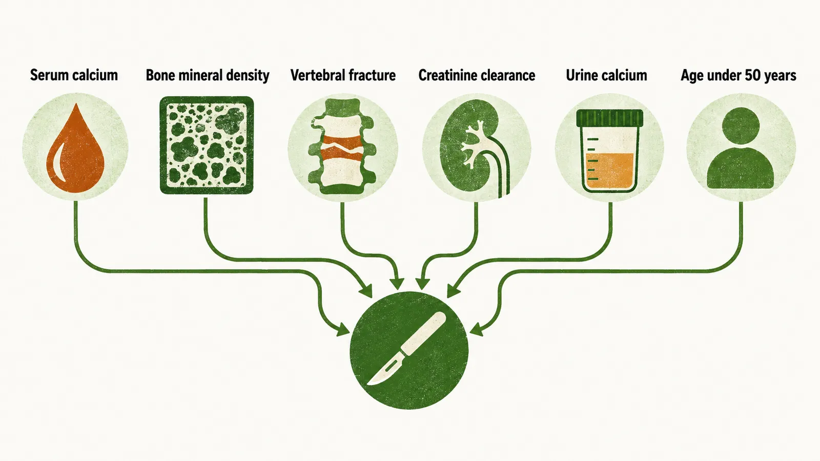
- Serum calcium more than 1 mg/dl above the ULN.
- Bone mineral density by DEXA T-score ≤ -2.5, in each of the lumbar spine, total hip, femoral neck, or distal third of the radius. This measures the bone loss PTH causes.
- Vertebral fracture on imaging by X-ray, CT, MRI, or vertebral fracture assessment, which shows that bone has already failed.
- Creatinine clearance < 60 cc/min, which means the kidney is already impaired.
- 24-hour urine calcium > 400 mg/day together with an increased biochemical stone risk profile. High urine calcium and crystal risk lead to stones.
- Nephrolithiasis or nephrocalcinosis found by ultrasound, CT, or X-ray, which is the kidney damage the clinical presentation note describes.
- Age under 50 years.
What if surgery is not chosen?
Some patients do not meet the criteria or do not want surgery, so medical management is used. Patients who meet none of these conditions, or who cannot have or decline surgery, are managed medically. Medical management keeps calcium and vitamin D at normal levels.
Where hypercalcemia or bone loss needs treating, a calcimimetic such as cinacalcet lowers PTH and serum calcium. Cinacalcet makes the CaSR more sensitive to calcium, so PTH is turned down at the same calcium. A bisphosphonate improves bone mineral density without correcting the hypercalcemia. Some bisphosphonates act on the bone-removing cells, so bone breakdown falls, but the gland is still the source of PTH, so calcium stays high.
What does the whole disease look like, from the first high calcium to the damage it leaves?