Primary hyperparathyroidism can appear in unusual settings. The setting changes how urgent the problem is, and it changes which other diseases must be ruled out.
How does the parathyroid gland read the calcium level?
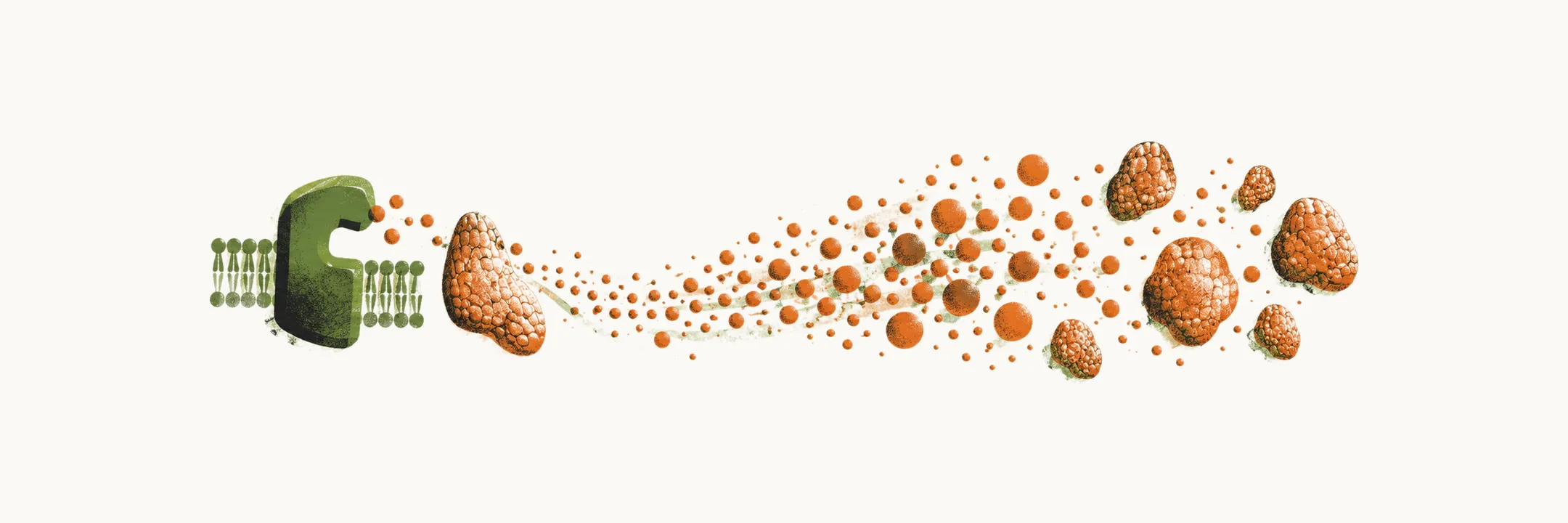
Each parathyroid chief cell carries a calcium-sensing receptor (CaSR) on its surface. The receptor reads how much calcium is in the blood around the cell, and the cell adjusts its PTH release accordingly. When calcium is high, the cell releases less PTH; when calcium is low, it releases more. The receptor controls PTH secretion tightly across the range of calcium it senses.
An unusual form of primary hyperparathyroidism is a place where this control breaks, is set differently, or where the setting changes what matters: a gene that switches the sensor off, a mother’s calcium reaching her baby, a crisis, a carcinoma, or cells scattered during surgery.
What goes wrong when the sensor is missing in newborns?
The neonatal forms of primary hyperparathyroidism are rare, and they are genetic. They are associated with the CaSR, and they can come from inactivating changes in the gene for the sensor.
The sensor is the brake on PTH release, so what happens when the brake is absent? With a homozygous mutation, both gene copies are affected. The parathyroid cells then release PTH in an excessive, unregulated way. PTH keeps flowing whatever the calcium level, so calcium rises to extreme levels. The result is severe and fatal hypercalcemia.
What about a single changed copy?
If both copies must fail for the newborn picture, what does one changed copy do? With a heterozygous mutation, only one copy is affected, and the hypercalcemia is mild and benign. This mild form is also known as familial hypocalciuric hypercalcemia (FHH).
What does maternal calcium do to the fetus?
A lifelong mild form is one end of the range; a mother with primary hyperparathyroidism affects her baby in a different way. Primary hyperparathyroidism in pregnancy raises concerns about the fetus and the neonate. The commonly reported effects are spontaneous abortion, low birth weight and supravalvular aortic stenosis.
There is also a risk to the baby after birth. The mother’s high calcium crosses the placenta, so the fetal parathyroid glands are exposed to a lot of calcium during fetal life, and this suppresses them.
Why does the newborn’s calcium fall after birth?
The fetal glands were suppressed in the womb. What happens when that calcium supply stops? The newborn is accustomed to the suppressed state, so the glands respond inappropriately to the falling calcium. The resulting hypoparathyroidism leads to tetany and hypocalcemia during the first days of life. Neonatal tetany is therefore another risk of primary hyperparathyroidism in pregnancy.
What is a parathyroid crisis?
Pregnancy changes who is affected; a crisis changes how fast the damage happens. Acute primary hyperparathyroidism is a sudden, life-threatening episode of hypercalcemia in a patient with primary hyperparathyroidism. It is also called parathyroid crisis, parathyroid poisoning, parathyroid intoxication or parathyroid storm.
Several organs are often affected at once, which is why the crisis is life-threatening. Commonly the serum calcium is very high, and the PTH concentration is high too, up to 20-fold.
What does the crisis do to the kidneys and bones?
The organ effects come from the same excess calcium and PTH that earlier notes explained. A crisis commonly shows nephrolithiasis and radiological evidence of subperiosteal bone resorption.
Why can the crisis look like a carcinoma?
The laboratory picture of a crisis resembles that of parathyroid carcinoma. Crisis bloodwork can look like a cancer, so what is a parathyroid carcinoma?
What is parathyroid carcinoma, and what sets it apart?
Parathyroid carcinoma accounts for 0.5% of primary hyperparathyroidism. It is the cancer the crisis may mimic. How is it linked genetically? It is strongly associated with hyperparathyroidism-jaw tumour syndrome (HPT-JT), a rare autosomal dominant condition, and carcinoma occurs in 15% of HPT-JT cases. The carcinoma and the syndrome share a gene: the gene behind HPT-JT is also altered in most sporadic carcinomas.
Carcinoma can be told from benign primary hyperparathyroidism by a far greater increase of serum calcium and PTH. The overlap is real, though. Average levels are higher in carcinoma than in adenoma, but most severe elevations are still benign, so bloodwork alone cannot decide.
How does a carcinoma show itself?
The clinical manifestations are these:
- nephrolithiasis in 60%
- skeletal involvement on radiological imaging in 35-90%
- a palpable neck mass in 30-76%
The stones and the bone findings are the same kidney and bone effects of excess calcium and PTH met earlier. The neck, kidney and bone findings lead on to where the carcinoma spreads.
Where does a carcinoma spread?
Spread is one way carcinoma differs from benign disease. A parathyroid carcinoma can present with metastasis at a later stage. The metastases are located mostly in the lung, the liver and the lymph nodes.
What about MEN1?
Parathyroid carcinoma is only very rarely reported in MEN1 syndrome. In MEN1 syndrome the parathyroid tumours are almost always benign.
What happens when parathyroid tissue is scattered?
A carcinoma is one tumour in one place; parathyromatosis is many small nodules, most often made by seeding. It is characterised by nodules of parathyroid-derived tissue embedded in the soft tissues around the gland. Parathyroid tissue spilled during surgery seeds the soft tissue of the neck and grows into nodules. The majority of reported cases are in patients who had parathyroid surgery for secondary hyperparathyroidism, and those patients had end-stage renal disease.
So how do you tell these unusual forms from the usual disease, and what is done first?