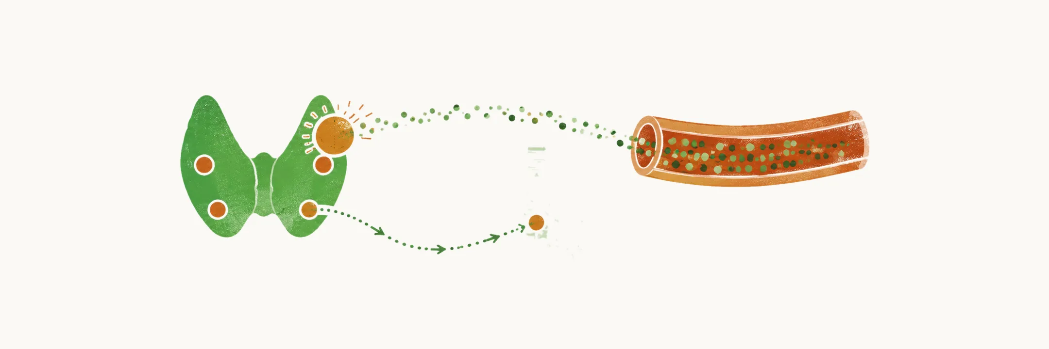
Primary hyperparathyroidism (PHPT) is over-production of PTH by one or more parathyroid glands. Who develops the disease, how it was found and how it arises all shape how it presents.
The parathyroid glands make PTH, which raises blood calcium when calcium falls, by acting on bone, the kidney and the gut. A normal gland senses calcium through the calcium-sensing receptor on its surface and makes less PTH as calcium rises, so calcium stays in a narrow range. In PHPT that control is lost: the gland keeps making PTH even when calcium is high, so the calcium stays high and both the excess hormone and the excess calcium act on the body.
Where are the parathyroid glands?
PHPT is a gland making too much hormone, so the first thing to know is where the glands sit.
The parathyroid glands normally sit as two pairs on the back of the thyroid gland. They end up there because the lower glands stop on the thyroid after moving down in the embryo.
They can also be found in ectopic places: within the thyroid gland, in the mediastinum, or within the thymus gland. This happens because the thymus and the parathyroid glands travel down into the neck together in the embryo, so a gland can be left with the thymus or carried further down into the chest.
An ectopic gland matters clinically because it can be missed during parathyroidectomy. A gland that sits off the usual site can be missed when the operation is done at the usual site.
How does a gland come to make too much PTH?
Knowing where the glands sit explains where a stray gland can hide; the next question is what makes a gland overproduce.
PHPT is caused by over-production of PTH from one or more parathyroid glands. The sensing point of the gland is shifted, so PTH keeps coming when calcium is already high. In most cases the etiological cause is not known, and the disease occurs sporadically.
What raises the risk?
In most cases no cause is found, but some exposures are known to raise the risk. These are factors for a higher incidence of PHPT:
- lithium use, which is reported to shift the same calcium-PTH set point, so that PTH is released at higher calcium levels
- external neck radiation
- chronically low calcium intake
- high body weight
Is the excess usually from one gland or several?
The risk factors raise the chance that a gland starts; the next question is how many glands take part.
A solitary parathyroid adenoma is by far the most common finding in PHPT, in more than 80% of cases. The excess then comes from one enlarged gland among normal ones.
In the remaining cases more than one gland is involved. This changes the surgical plan, because removing one gland leaves the others still overproducing, so the operation has to cover all of them.
What makes several glands involved?
Inherited syndromes are one reason more than one gland can grow. The risk factors for multiple gland involvement are:
- MEN1 (multiple endocrine neoplasia type 1)
- MEN2 (multiple endocrine neoplasia type 2)
- lithium exposure
MEN1 is inherited, so the altered gene is present in the body’s cells from the start. A second change in a gland can then let that gland grow, which is consistent with Knudson’s two-hit model of hereditary cancer. MEN2 is also inherited, and in MEN2 syndromes several parathyroid glands can be involved as adenomas.
Who develops PHPT?
With the glands and risks in place, the next question is who actually gets the disease.
PHPT is a disease of middle-aged people, mostly diagnosed between 50 and 60 years of age. It is more common in women, with a ratio of 3-4:1. Recent studies show that it is also more common in African-American people.
How did PHPT look before automated testing?
Who gets it, and how it arises, shaped how it looked; the older picture started in the kidneys.
In the past PHPT presented with nephrolithiasis. The kidney filters a large amount of calcium, and PTH helps it hold calcium back. When blood calcium is high, the filtered load is larger than the kidney can reclaim, so urine calcium runs high, and stones can form.
What did it do to the bones?
The same high calcium and PTH also reached the skeleton. Sustained PTH excess drives osteoclasts to break down bone, and fibrous tissue replaces the bone in cystic spaces. This is osteitis fibrosa cystica. PTH-driven osteoclasts also remove bone just beneath the periosteum, so the bone surface is cut away from underneath. This is sub-periosteal bone resorption.
Two more changes were also part of the past picture: distal tapering of the clavicle and phalanges, and salt-and-pepper erosions of the skull.
What did it do to nerves and muscles?
The skeleton was not the only tissue affected. The past presentation also included neuromuscular dysfunction and weakness.
What did the case series show?
The older picture is visible across case series, which show the shift. In the table, NR means not reported.
| Series | Nephrolithiasis | Skeletal disease | Hypercalciuria | Asymptomatic |
|---|---|---|---|---|
| Cope (1930-1965) | 57% | 23% | NR | 0.6% |
| Heath et al (1965-1974) | 51% | 10% | 36% | 18% |
| Mallette et al (1965-1972) | 37% | 14% | 40% | 22% |
| Silverberg et al (1984-2009) | 17% | 1.4% | 39% | 80% |
What changed with automated calcium testing?
The series show complications falling, which fits the change in how the disease was found.
The change came with automated laboratory techniques for detecting and measuring hypercalcemia in the 1970s. Calcium is now measured routinely, so a rise in calcium is found before it causes symptoms or damage. Since then more than three fourths of patients with PHPT are diagnosed in the asymptomatic stage. The fall of major complications across the successive case series shows this shift. PHPT is nowadays not seen as severely as in the past.
Can PHPT be found with a normal calcium?
Once testing finds the disease early, the next question is whether it can be there when calcium reads normal.
In the 1990s, screening for osteoporosis in postmenopausal women was introduced, and this led to another diagnostic breakthrough. It became clear that it is possible to have normocalcemia with a high level of PTH. Because the sensing point is shifted, PTH can run inappropriately high for a calcium that reads normal. This led to a new entity called normocalcemic PHPT, or NPHPT.
Once the calcium is high, what does that do to the rest of the body?