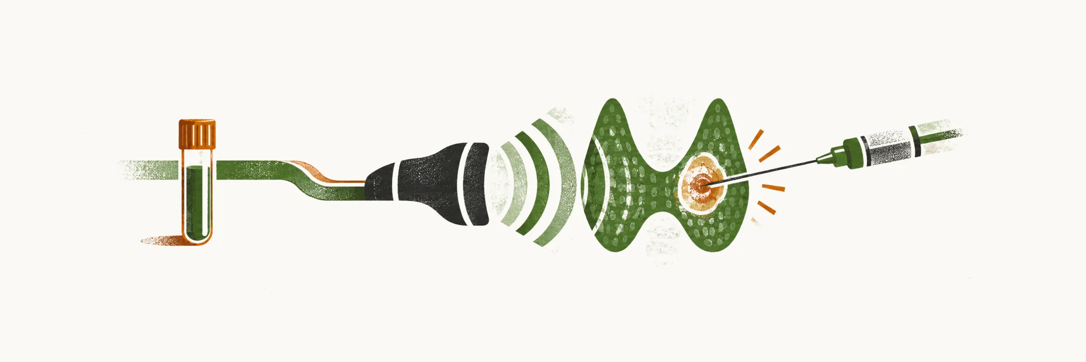
A nodule has been found, and the work-up asks one question: is it cancer? Most nodules are benign, as the multinodular goiter note showed, so the work-up exists to find the few that matter without sampling every nodule.
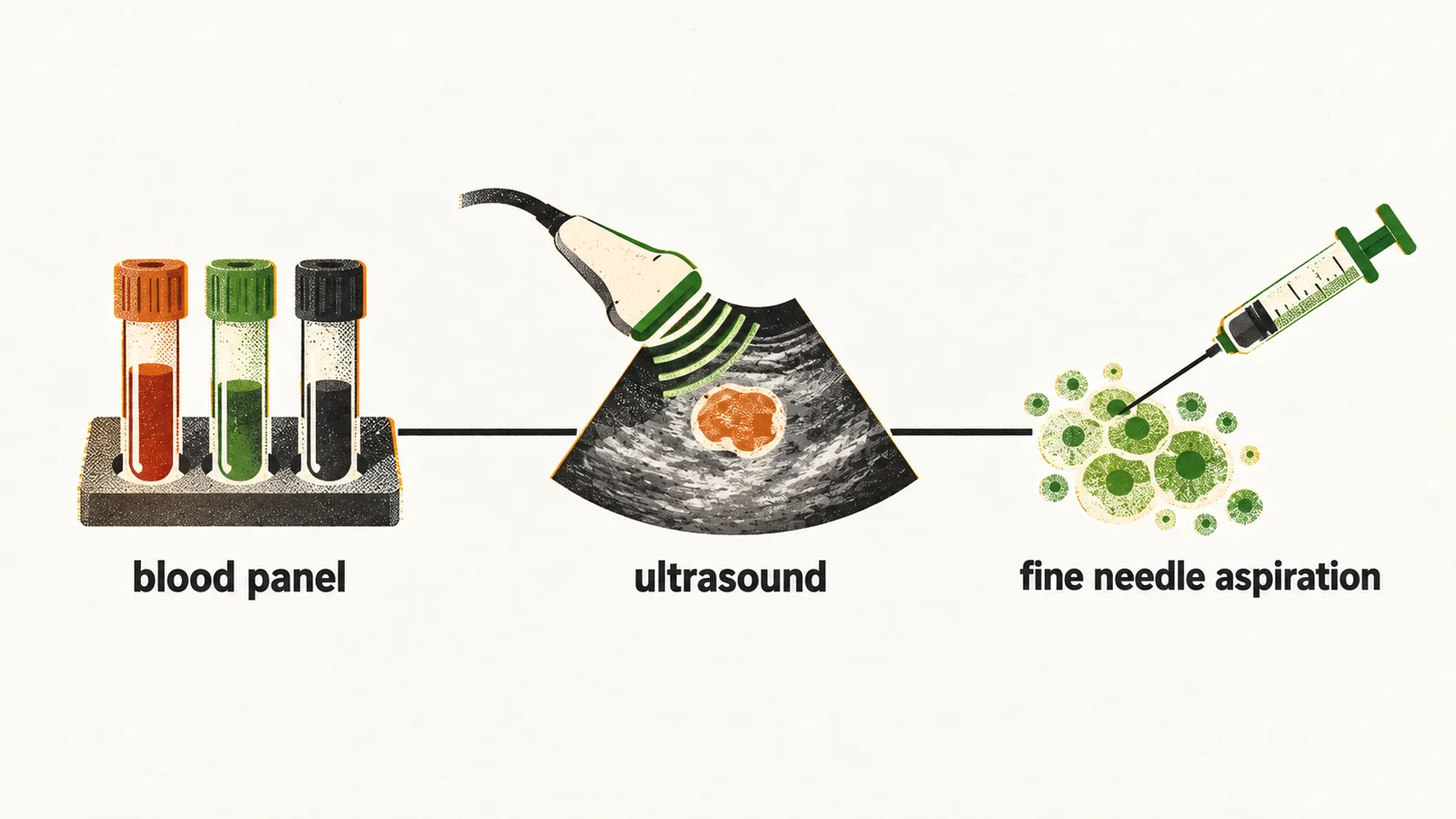
Diagnosis rests on three tests: biochemical tests, ultrasound imaging and fine needle aspiration. They come in that order, and each asks something the others cannot. The blood panel checks whether the gland is working in some other way, and whether the immune system or a calcitonin-making tumour is involved. The ultrasound shows how the nodule looks, and that look estimates how likely cancer is. The needle reads the cells themselves, and it is used when the look makes sampling worthwhile. Each step narrows what the next one has to answer.
Where does the work-up start?
It starts with the blood panel. Its purpose is mainly to rule out benign conditions. The measured parameters should cover thyroid function, malignancy and autoimmune thyroid disease.
The hormone results come first, so what do they show?
What do the hormone results say?
The panel includes TSH, T4 and T3. The pituitary’s TSH drives the gland, as the goiter note showed, so a low TSH can mean that a nodule is working without waiting for that signal. T4 and T3 are the hormones the gland releases, and their rise is what turns TSH down, as the toxic nodule note showed.
The results can point to other causes of nodules. A normal TSH with a normal calcitonin points to a non-toxic multinodular goiter, and a normal TSH fits nodules that do not make hormone on their own, as the multinodular goiter note showed. A low TSH with raised thyroid hormones and a normal calcitonin points to a toxic multinodular goiter, because a nodule that makes hormone on its own shows high hormone and low TSH in the blood.
If the hormones are not the explanation, is the immune system involved?
Is the immune system attacking the gland?
The next tests are antibodies. Antibodies against thyroid peroxidase (TPO) are the immune system attacking the gland, and such antibodies damage the gland. Antibodies against thyroglobulin (Tg) are the same kind of attack, aimed at thyroglobulin instead. Thyroglobulin is a protein made by follicular cells, as the cancer note explained, and its level in serum is also part of the panel.
Elevated antibodies mean the immune system is inflaming the gland. So positive thyroid antibodies point to autoimmune thyroid disease.
A second marker looks for a different cell type, so what does calcitonin add?
Could it be a calcitonin-making tumour?
Calcitonin is made by C cells, and medullary thyroid carcinoma grows from C cells. A raised calcitonin therefore points to that tumour, and the test is used for screening for it.
Still, measuring calcitonin in every nodule is not firmly recommended. One reason is false positives. A high calcitonin can come from benign tissue with C-cell hyperplasia around a nodule, so a raised value does not always mean cancer. The other reason is that the benefit of routine measurement is uncertain.
The blood tests cannot settle the nodule itself, so what does the nodule look like on ultrasound?
What does the nodule look like on ultrasound?
The panel mainly rules out benign causes, so the question of cancer falls to the nodule. Ultrasound comes before the needle, and the American Thyroid Association guideline for fine needle aspiration is therefore based on ultrasound findings. Whether a nodule is biopsied depends on its size and its ultrasound pattern, as the multinodular goiter note showed.
Some features carry more weight than others. Microcalcifications and irregular margins are the strongest predictors of malignancy. A cancer that grows into the tissue around it does not have a clean edge, as the cancer note showed. Microcalcifications are also seen in some benign lesions, so they raise suspicion rather than settle it.
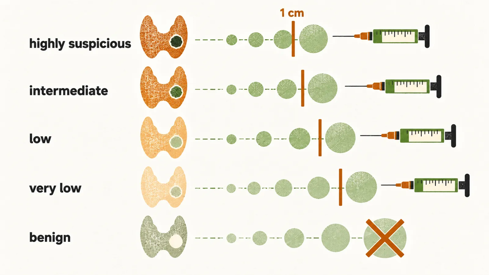
The pattern and the size together set the threshold for sampling, and the more suspicious the pattern, the smaller the nodule that is sampled:
- A highly suspicious pattern is sampled from 1 cm upward.
- An intermediate-suspicion pattern is sampled from 1 cm upward. A smooth, solid, hypoechoic nodule lacks the strongest features, so it sits one step lower on the scale.
- A low-suspicion pattern is sampled from 1.5 cm upward. A solid isoechoic or hyperechoic nodule lacks the strongest features, so the threshold is set later.
- A very-low-suspicion pattern is sampled from 2 cm upward. Nodules that are mostly cyst look spongiform on ultrasound, as the multinodular goiter note showed.
- A benign pattern is not sampled.
When the look meets the threshold, what does the needle add?
What does the needle add?
A thin needle goes into the nodule and brings back its cells, which are read under the microscope.
What is done with the result follows from the behaviour and risk of the nodule: observation for a benign nodule, surgery and further treatment for cancer. It does not follow from the biopsy alone. Some results, such as suspicious cytology or a follicular neoplasm, are less certain.
Once the needle has placed the nodule in a risk group, what decides whether it is watched, removed or treated?