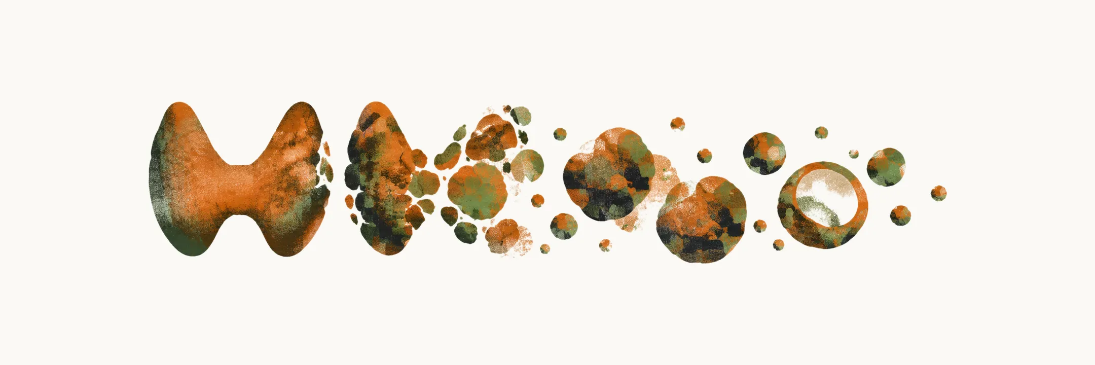
Multinodular goiter (MNG) is a thyroid that enlarges by forming several nodules instead of growing evenly. The thyroid sits in the front of the lower neck, with its two lobes against the sides of the windpipe. The gullet lies just behind the windpipe, and large neck veins run beside the gland. There is little room to spare.
What does a goiter made of nodules look like?
The gland can grow evenly, or in patches, as the diffuse goiter note showed. In MNG it grows in patches.
Molecular studies support this. The cells of different nodules are clonally different. This suggests hyperplasia in response to local growth factors and cytokines, and it argues against the spread of a single clone.
Nodules may be solitary or multiple, and functioning or non-functioning. They also do not always enlarge the gland. Discrete nodules can be present in a thyroid of completely normal size.
Why does the gland grow in lumps, and where is it most common?
MNG occurs mostly in regions with iodine deficiency. Iodine shortage keeps TSH high, and that keeps the follicles growing, as the diffuse goiter note explained. MNG can also be seen in iodine-sufficient areas. There it is due to genetic, autoimmune and environmental effects. These causes are several and can combine, so no single cause explains every case.
The next question is what the nodules are made of.
What are the nodules made of?
Nodules are of two broad forms, hyperplasia-related and neoplastic-related, and two forms of benign nodule are found in the thyroid gland.
The histological features of nodules are quite variable. A nodule may show:
- hypercellular areas;
- cystic areas filled with colloid;
- extensive fibrosis;
- haemorrhage;
- lymphocyte infiltration.
Hyperplastic nodules reflect both micro- and macrofollicular architecture. They mostly appear as a combination of cysts and cells. On ultrasound they mostly show a spongiform pattern, in which more than 50% of the nodule is cysts.
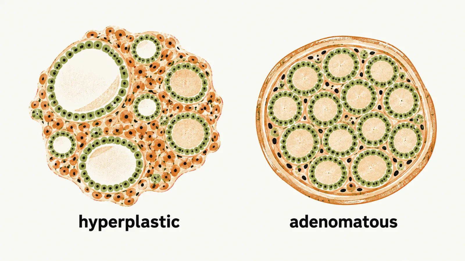
Adenomatous nodules are mostly monotonous, microfollicular, encapsulated nodules.
Are these nodules dangerous?
Most of the lumps are benign, so the real question is which ones cause trouble. Thyroid nodules are very common in adults. A nodular finding is felt on neck examination in about 3-7% of adults. Up to 50% of adults carry nodules smaller than 1 cm, and these are found only by ultrasound.
Fewer than 7% of nodules found by both palpation and neck ultrasound are malignant. More than 93% are benign lesions. Likewise, the majority of solid thyroid nodules are benign.
Most patients with non-toxic MNG are asymptomatic. Most are also euthyroid. The change develops over several years. MNG is mostly an incidental finding, noticed by the patient or by the clinician during physical examination or imaging.
What does a large gland do to the neck?
The neck has little room, so a growing gland presses on what lies beside it. Compressive effects appear only as very uncommon manifestations. They occur when a large enough goiter presses on peripheral structures. Each structure gives its own symptom:
- Obstruction of venous outflow causes venous congestion. Blood backs up behind the pressure, so the face and neck look congested.
- Tracheal compression causes respiratory distress (dyspnoea), because the narrowed windpipe makes breathing harder.
- Oesophageal compression causes dysphagia, because the narrowed gullet makes swallowing hard.
- Hoarseness of voice reflects laryngeal nerve involvement. Hoarseness is specific for malignant forms of MNG.
Sudden pain in MNG is due to haemorrhage inside a nodule.
On physical examination the different nodules produce a distorted thyroid gland. Some nodules are not appreciable because of their location, for example posterior or substernal nodules. A nodule behind or below the sternum lies inside the chest, where the fingers cannot reach.
Is there a bedside sign?
In MNG with a substantial goiter, Pemberton’s sign can be elicited. The patient raises both hands, and cyanosis of the face appears. It is due to an increase of pressure in the upper thoracic inlet.
What tests show how far the goiter has gone?
The tests measure what the sign suggests, and the TSH level says something about the lumps. In patients with these signs and symptoms, a few functional and biochemical values should be checked. The TSH level is checked mainly to exclude thyroid dysfunction. TSH rises when the gland makes too little hormone, as the diffuse goiter note showed. In MNG the TSH level is mostly normal.
Other tests show the extent of the problem:
- Pulmonary function tests show how much tracheal compression is present.
- CT or MRI show the amount of enlargement, the extent of substernal extension, and the degree of tracheal narrowing or oesophageal compression.
Whether a nodule in MNG is risk-stratified and biopsied depends on its size and its sonographic pattern. ACR TI-RADS (American College of Radiology Thyroid Imaging Reporting and Data System) is used for that stratification.
Does a low TSH tell us anything about the lumps?
Because the TSH level is mostly normal in MNG, a low TSH in a nodular gland points to nodules that make hormone on their own. This fits the fact that nodules may be functioning or non-functioning. But what happens when one of these nodules starts making hormone without waiting for TSH?