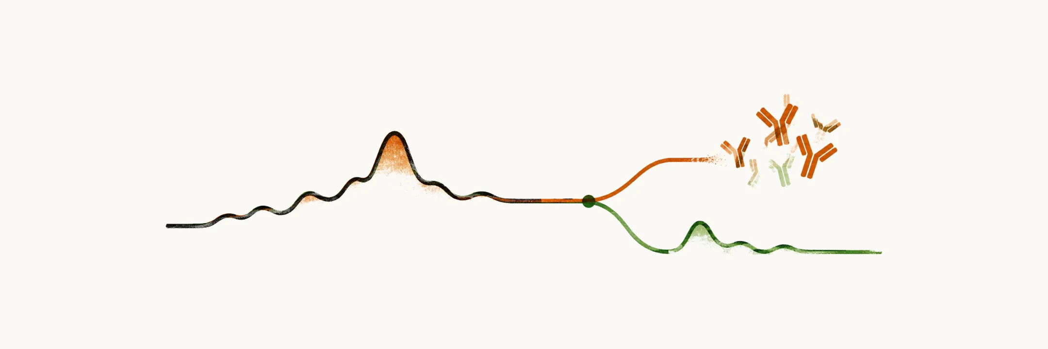
Diagnosing T1D takes two steps, in a fixed order. The first asks whether the glucose is high enough to be diabetes at all. The second asks what has made it high. Insulin moves glucose from the blood into cells, so when the insulin supply fails, glucose stays in the blood. In T1D the immune system has destroyed the beta cells that make insulin. The tests of the second step therefore look for that destruction and for how much insulin the cells still make.
Is there diabetes at all?
The first step shows that the patient has diabetes. It uses the same glucose thresholds that define every form of the disease. The ADA criteria define diabetes by any one of four measurements:
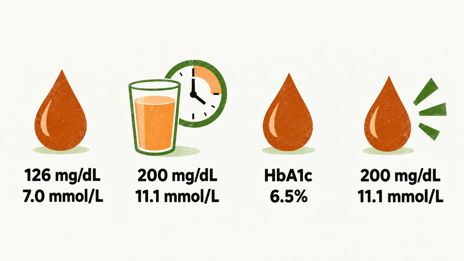
- fasting plasma glucose of 126 mg/dL (7.0 mmol/L) or more;
- 2-hour plasma glucose of 200 mg/dL (11.1 mmol/L) or more during a 75-gram oral glucose tolerance test;
- HbA1c of 6.5% or more;
- random plasma glucose of 200 mg/dL (11.1 mmol/L) or more in a person with classic symptoms of hyperglycemia or a hyperglycemic crisis.
The presentation note explained why the classic symptoms come from the high glucose itself. So in a clearly symptomatic patient, a single random glucose at or above 200 mg/dL is enough to confirm diabetes.
When the hyperglycemia is not unequivocal, a second abnormal result confirms the diagnosis. It may come from the same sample or from a repeat sample on another day. The values behind these thresholds are set out in detail in Diagnostic Criteria for Diabetes and Prediabetes.
These thresholds cannot name the kind of diabetes. Type 1 and type 2 both leave too little insulin for the glucose, so the same high glucose can come from either. Glucose shows that diabetes is present, not which kind.
If so, what is causing it?
The high glucose settles that diabetes is present, but not why the insulin has failed. Once diabetes is confirmed, the next question is which kind, because treatment differs.
That is the second step: it shows that the cause is autoimmune destruction of pancreatic beta cells. T1D is defined as diabetes caused by this destruction, and the destruction is what leads to absolute insulin deficiency. The cause also decides what is left of the insulin supply. So this step separates T1D from type 2 diabetes, where the cells still secrete, and from its slower adult variant, where the loss is slow.
How much insulin is left?
Step 2 shows the cause is autoimmune destruction, and destruction leaves a particular kind of shortage. Beta cells are the source of insulin, so when they are destroyed the insulin is absent rather than reduced. Autoimmune destruction usually leads to absolute insulin deficiency.
This is the first and most important difference between T1D and type 2 diabetes: the insulin secretion deficiency is absolute in T1D and relative in type 2. The difference is whether any insulin is still made. In type 2 the beta cells still secrete insulin, just not enough for the degree of insulin resistance. The cells first secrete more to match the resistance, and diabetes appears when they cannot keep up. So the shortfall is relative, not absolute.
What do the antibodies show?
A shortage of insulin alone cannot name the kind. Two laboratory markers do most of the separating, and the first is islet autoantibodies. The autoantibodies are the visible trace of the immune attack on the beta cells, as the natural-history note set out. Their presence shows the immune system is attacking the beta cells.
The main islet autoantibodies are:
- antibodies to glutamic acid decarboxylase (GAD), the most commonly found;
- antibodies to tyrosine phosphatase (IA-2);
- antibodies to a zinc transporter (ZnT8);
- antibodies to insulin (IAA);
- islet cell antibodies (ICA).
At least one islet autoantibody is present in the great majority of people with newly diagnosed T1D. A small group with T1D has none of these antibodies. A test panel can miss autoimmunity, so the absence of islet autoantibodies does not exclude T1D.
How much insulin does the pancreas still make?
The antibodies show the attack, but not how much insulin the cells still make. C-peptide does. It is released together with the cells’ own insulin, so it measures what the cells still make, as the natural-history note set out. It measures the patient’s own insulin output, not injected insulin.
A low or undetectable C-peptide level means the cells are making little, which is what absolute insulin deficiency looks like. It therefore documents absolute insulin deficiency. In type 2 diabetes the cells keep secreting, so a low level also supports T1D over type 2 diabetes.
What do ketones add?
An insulin shortage can show in the blood as ketones. As the presentation note showed, when insulin is too short the body breaks down fat and the liver makes ketone bodies. Ketones in blood or urine therefore point toward T1D. Relative lack of insulin can do the same, so ketones point toward T1D without proving it.
Frank ketoacidosis at presentation is the same mechanism at its most advanced, so it points the same way.
What if the picture looks like type 2?
The markers so far separate T1D from type 2 by what is left and what caused the loss. Some adults have an autoimmune cause with a slow course, and that is the hard case. The name for this case is latent autoimmune diabetes in adults (LADA).
LADA has a mild autoimmune process, so beta-cell destruction progresses more slowly than in classic T1D, and beta-cell function falls faster than in type 2. Because the loss is slow, residual insulin lasts, and LADA patients may not need insulin for months or years. Slow progression and an adult course resemble type 2 diabetes, so LADA patients are easily mistaken for having type 2 diabetes.
The cause is autoimmune, so islet autoantibodies are what reveal LADA. The antibodies most often found in LADA are GAD antibodies.
What does an absolute shortage mean for treatment?
If the autoimmune loss is complete, insulin cannot be relied on from the body. With no residual supply, the body has nothing to fall back on, so insulin is often needed from the start. As the epidemiology note set out, once the beta cells are gone the body cannot make insulin for life, so it has to come from outside. Absolute deficiency means insulin will eventually be needed to survive. The route to the Treatment of type 1 diabetes starts here.
If the pancreas can never make enough insulin again, what supplies the insulin the body needs every day?