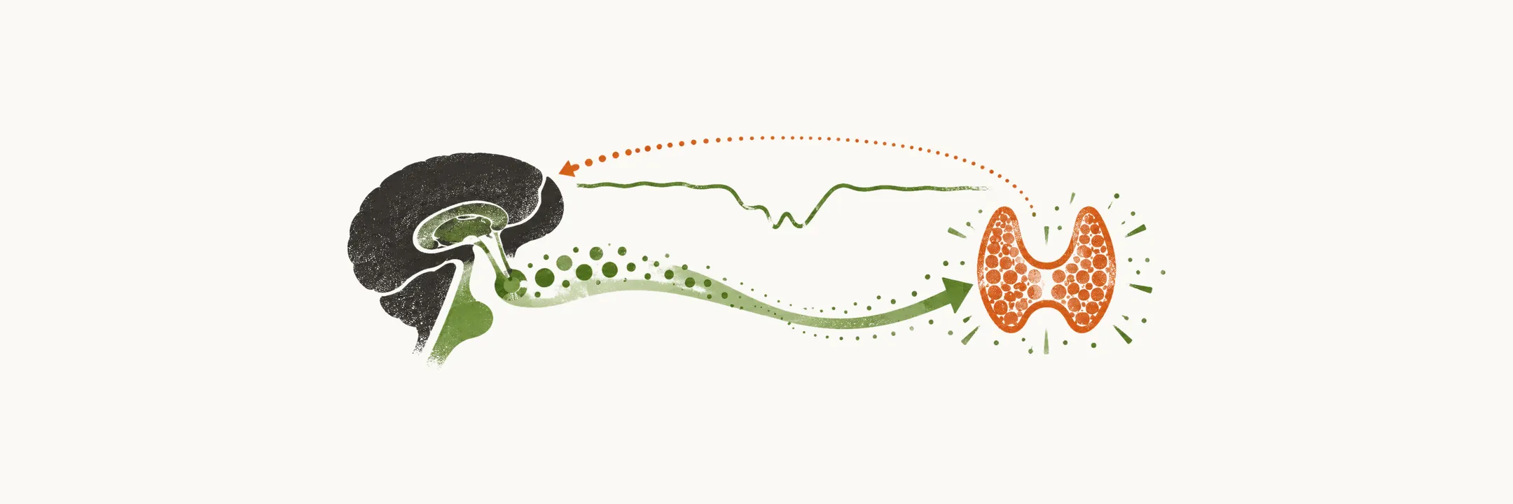
Which blood test comes first when hypothyroidism is suspected, and what does each further test add? Laboratory testing starts with TSH. The main evaluation is TSH together with free T4.
The picture is a loop. The pituitary watches the blood for thyroid hormone and sends TSH to push the gland when the hormone falls. The link between the two is steep, so a small fall in hormone brings a large rise in TSH. Each test looks at a different part of that loop.
Why does the first test measure TSH?
Testing starts with TSH because the pituitary’s response to a small fall in thyroid hormone is large, so TSH shows the change first. The change in TSH is much larger than the change in the hormone itself. In figures, a 2-fold change in T4 level is associated with up-to-100-fold change in TSH.
This amplification makes the TSH assay far more sensitive. A small loss of gland output shows up as a swing in TSH that a routine laboratory reads easily. That is why TSH is the gold standard test.
Because TSH moves first and far, it becomes recognizably abnormal long before T4 falls outside its reference range. TSH is therefore measured first, and not T4.
What does a free T4 result add?
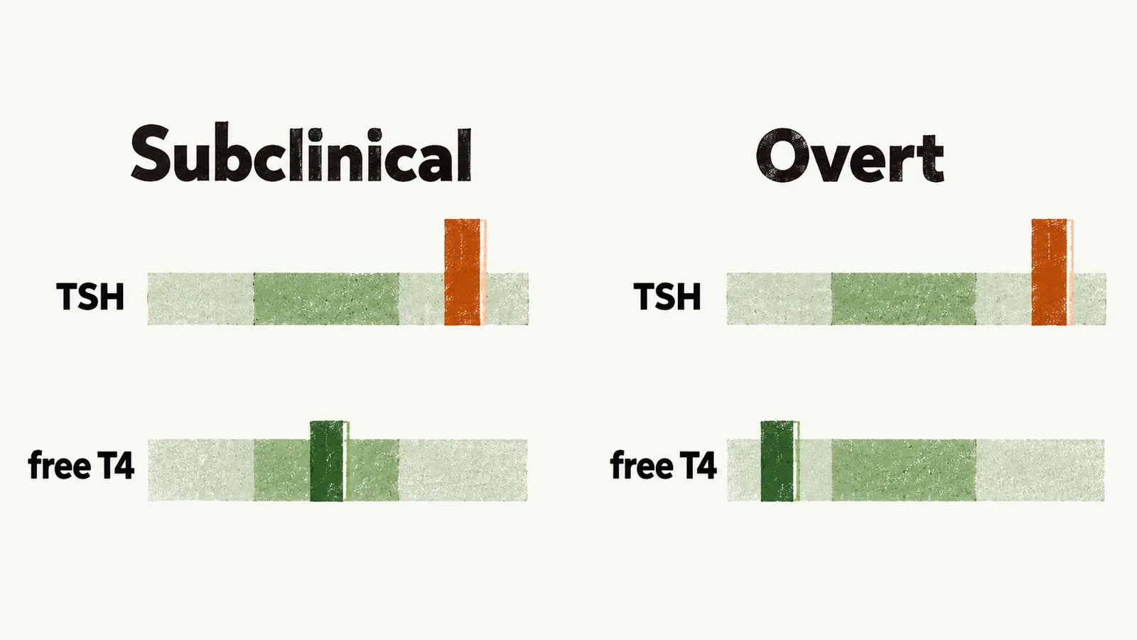
TSH shows that the pituitary is pushing. The second test asks whether the hormone itself has fallen. If TSH is increased, the diagnosis needs confirmation by free T4 level testing.
Free T4 is not the first test because the hormone stays in range for a time while the pituitary still compensates. In subclinical hypothyroidism free T4 is normal, as the subclinical note showed. Free T4 is done after a raised TSH to confirm the diagnosis, and it separates subclinical from overt disease: overt disease is where free T4 has fallen, once the reserve is used up.
What if the fault is in the pituitary?
The same loop can break at the pituitary, and then TSH stops being the guide. TSH is unreliable when the problem is central rather than in the gland, because the pituitary cannot raise it. This is why the causes note showed TSH stops being a reliable gauge there.
Why is T3 left out of the routine?
Free T4 measures the hormone that the gland makes. T3 is different, because the tissues help hold it steady. Much of T3 is made in the tissues by converting T4, so the gland’s output is only part of it.
Deiodinase activity increases as a compensatory mechanism and converts more T4 into active T3. T3 level is within normal range in more than 25% of cases. It is often normal even when hypothyroidism is significant.
So a normal T3 says little about how much the gland is making, and T3 is not used in the usual clinical investigation.
What do the antibody tests add?
Once the thyroid tests show hypothyroidism, the question becomes why the gland is failing. Antibody immunoassays are done once clinical or subclinical hypothyroidism is suspected and confirmed by TSH and free T4 levels.
They look for antibodies against TPO and against Tg, and they show whether the cause is autoimmune. TPO and thyroglobulin are the gland’s own proteins, as the autoimmune note explained. The antibodies mark the attack rather than start it, as the pathogenesis note explained.
TPO antibodies are the more useful of the two. They are found in about 90-95% of people with Hashimoto’s thyroiditis. Tg antibodies are found less often.
What else changes on the blood tests?
Low thyroid hormone slows many organ systems, so other blood tests can change too, as the clinical manifestations note showed. Beyond thyroid tests, other laboratory findings are abnormal in hypothyroidism:
- elevated creatine phosphokinase
- elevated cholesterol profile, especially LDL
- elevated triglyceride
- anemia
- hyponatremia
Muscle affected by low hormone leaks creatine kinase into the blood, because its cell membrane becomes more permeable, so the level rises. Low thyroid hormone raises LDL, the lipid change the subclinical note described, and triglyceride rises with the same lipid change.
Thyroid hormone acts on the kidney’s erythropoietin secretion. With less hormone, less erythropoietin is thought to be made, and red cell production is thought to fall. Effects on the marrow are also proposed contributors.
For hyponatremia, a proposed explanation runs through the kidney. Low hormone is thought to reduce cardiac output, which raises antidiuretic hormone and lowers filtration. Free water is then kept, and sodium is diluted.
Once the tests show the hormone is low and the cause is clear, what is given to replace it?