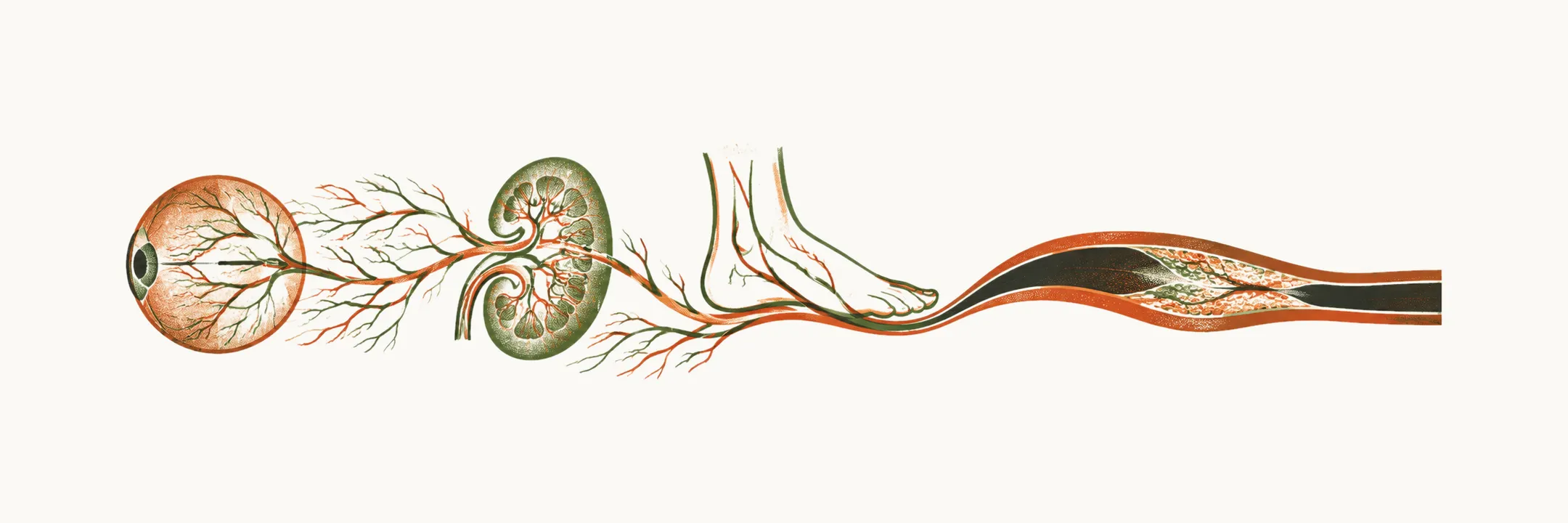
Persistent high glucose damages blood vessels, and that damage can already be present by the time the disease is recognised, as the presentation note explains. This vessel damage defines the long-term course of type 2 diabetes.
The complications are grouped into microvascular and macrovascular disease by the size of the vessels they affect. Microvascular disease affects the small vessels of the retina, the kidney and the peripheral nerves. Macrovascular disease affects the large arteries of the heart, the brain and the legs. Hyperglycemia often precedes the diagnosis by years, so some damage is already present when the disease is first recognised.
Why would the small vessels of three different organs all be hit?
Three small-vessel beds
Each of the three microvascular complications damages a different small-vessel bed: the retina, the kidney and the peripheral nerves. What does that damage look like first in the eye?
The eye
The first of the three is the eye. Diabetic retinopathy is damage to the capillaries of the retina. It is a leading cause of vision loss in working-age adults.
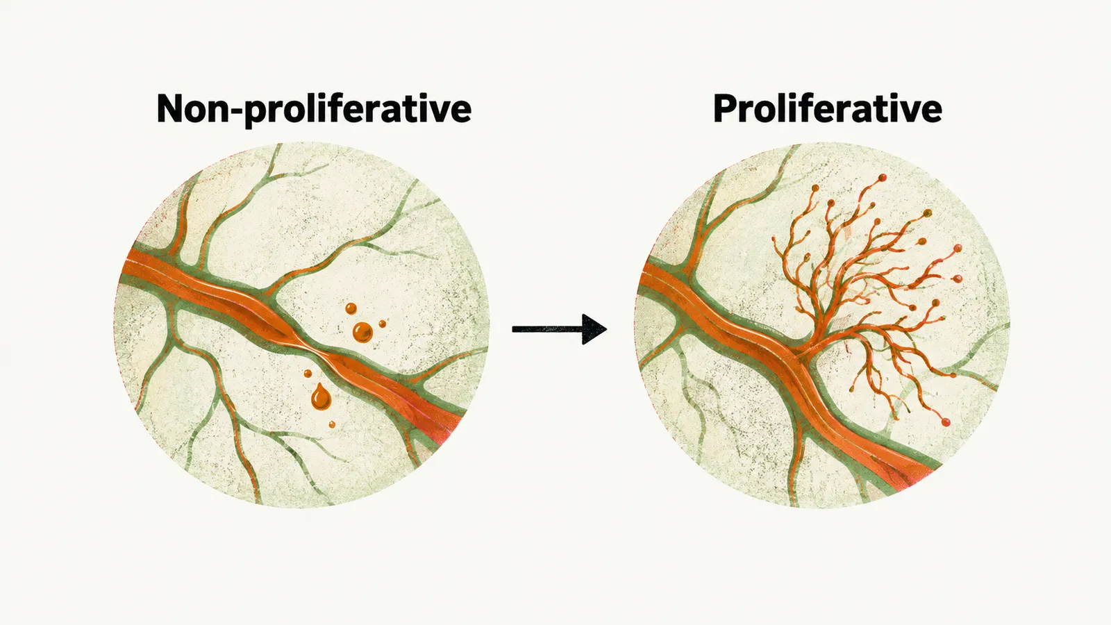
In the early non-proliferative stage, the vessels leak and occlude. This produces microaneurysms, hemorrhages and hard exudates.
In the later proliferative stage, hypoxia drives the growth of fragile new vessels. These vessels can bleed into the vitreous or pull the retina off, and the proliferative stage can cause severe visual loss.
The kidney is the second bed, so what does damage there do?
The kidney
The second bed is the kidney. Diabetic kidney disease is damage to the glomerular microvasculature. It begins with hyperfiltration. Clinically it then appears as albumin in the urine and a progressive fall in estimated glomerular filtration rate. Diabetic kidney disease is a leading cause of kidney failure.
The kidney takes back filtered glucose through SGLT2, and a proposed change in that transporter could keep glucose high; as the pathophysiology note explains, this is not yet firmly supported. In this proposed SGLT2 mal-adaptation, tubular cells increase their expression of SGLT2, which raises the renal reabsorption threshold for glucose. It might also add to the hyperglycemia behind the kidney damage.
The third bed is the nerves, so what is lost when they are damaged?
The nerves
The third bed is the nerves. Diabetic neuropathy is the most common of the diabetes-associated neuropathies, and it affects around half of people with diabetes.
The typical form is a distal, symmetric, sensory polyneuropathy of the feet. Because sensation is lost, injuries and pressure sores can go unnoticed. The autonomic nerves can be affected too, and autonomic neuropathy can involve the gut, blood pressure regulation and sexual function.
The large arteries are the other side of the damage, so what happens there?
The large arteries and the heart
In diabetes, atherosclerosis of the large arteries produces coronary artery disease, stroke and peripheral artery disease. Cardiovascular disease is the leading cause of death in people with type 2 diabetes.
Much of this cardiovascular risk is present before the glucose thresholds for diabetes are crossed. The reason is that the risk factors for diabetes and for cardiovascular disease overlap. Features that raise cardiovascular risk, such as high blood pressure and a high-triglyceride, low-HDL pattern, tend to appear in the same person, as the risk factors note explains.
The heart muscle itself is also affected. Heart muscle normally runs mostly on fatty acids and takes up glucose less well when fatty acids in the blood are raised, as the pathophysiology note explains. This myocardial insulin resistance means reduced glucose uptake by heart muscle, and it lowers the heart’s tolerance to ischemia.
The feet combine the nerve and artery damage, so what does that do to a wound?
The foot
The foot brings the nerve and artery damage together. Diabetic foot combines the mechanisms of neuropathy and of peripheral artery disease. Loss of protective sensation from neuropathy means that minor trauma is not felt. Peripheral artery disease reduces the blood supply that healing needs.
The result is that a wound becomes infected, fails to heal, and can progress to gangrene and amputation. A history of a diabetic foot event is associated with a marked increase in subsequent mortality.
All of this damage has a common thread, so is it preventable?
What the damage has in common, and how it is held back
The damage has one common thread: the complications are reduced by the same control that treats the disease. Good glycemic control lowers the risk of microvascular disease. Blood-pressure and lipid control carry much of the benefit against macrovascular disease.
Care is not organised around glucose alone. It is organised around periodic screening of the eyes, the kidneys and the feet.
If glucose, blood pressure and lipid control hold the damage back, what does a person change in daily life to reach that control?