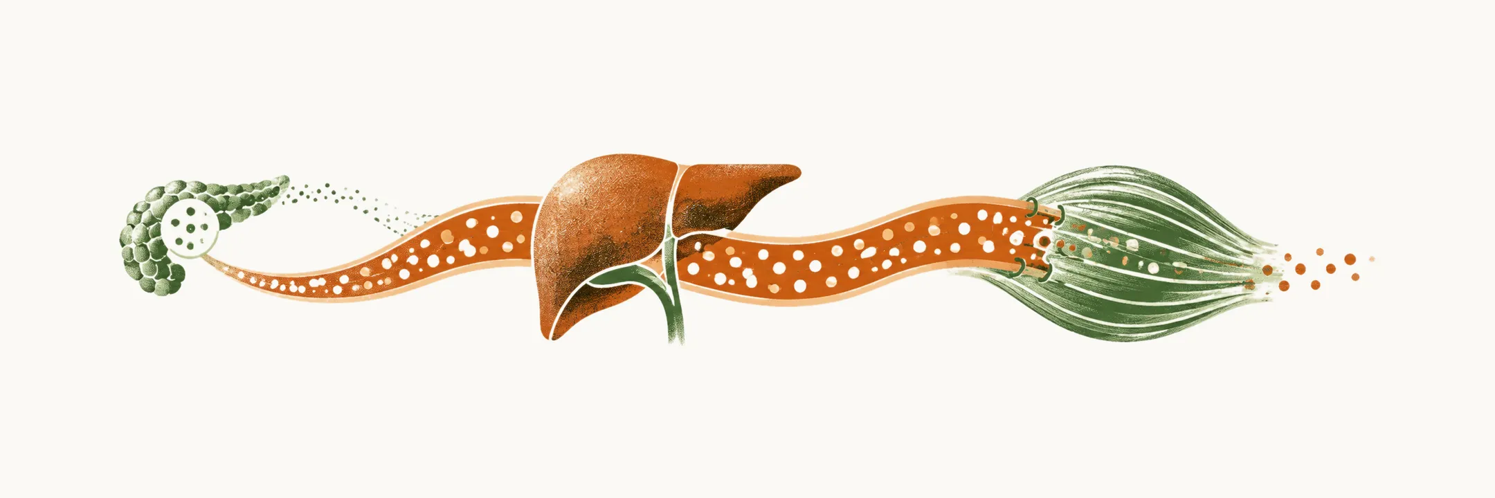
In T2DM the balance between insulin secretion and tissue sensitivity to insulin breaks down. Insulin is released into the portal vein, so the liver receives it first, before the rest of the body. Insulin tells the liver to hold back its glucose output, and it tells muscle and heart to take glucose up. Insulin resistance affects four glucose-handling sites: the liver, the kidney, skeletal muscle, and the heart and vessels. At each site the question is how tissue glucose handling departs from normal.
When these tissues respond less, glucose stays in the blood, and the β cells must release more insulin to hold it down. Normally, hyperinsulinemia and hyperglycemia augment glucose uptake, which reinforces tissue sensitivity. When tissues stop responding, that reinforcement is lost. As Normal glucose homeostasis shows, insulin sensitivity and β-cell function trade against each other, so a fall in one shifts the other. Insulin resistance is defined by two changes: reduced peripheral glucose uptake, mainly in muscle cells, and increased endogenous glucose production (EGP), the glucose the body itself releases into the blood, mostly from the liver. Adipocyte inflammation and adipocyte insulin resistance contribute. They bring increased lipolysis, a raised free fatty acid (FFA) level and increased intermediary lipid metabolites. These changes reduce peripheral glucose uptake further, increase endogenous glucose production, and make pancreatic β cells less sensitive to glucose.
Why would the liver be the first to show the failure?
Why the liver comes first
Physiologically, glucose ingestion makes the β cells secrete insulin into the portal vein. Because that insulin enters the portal circulation, the liver sees it before any other organ. Portal insulin inhibits basal EGP in the liver, and this prevents excessive hyperglycemia after a meal.
In T2DM the hepatic cells are insulin-resistant, so this inhibition is lost. Glucose then rises markedly after eating, because liver EGP is not suppressed by hyperinsulinemia. Fasting (post-absorptive) hyperglycemia has the same main mechanism, liver insulin resistance.
What does the liver do overnight?
What the liver does overnight
The size of this failure can be measured. In a study group of people with T2DM and mild-to-moderate fasting hyperglycemia, defined as glucose 140–200 mg/dL, night-sleep EGP is excessive, at about 2.5 mg/kg.min. Normal night-sleep EGP is 2 mg/kg.min. With no meal arriving overnight, most of the glucose comes from the liver’s output. It adds about 35 g of glucose by morning in an 80 kg individual.
The liver also responds steeply to insulin. The dose–response between EGP suppression and plasma insulin is very steep: only 30–40 µU/mL of insulin is enough to halve EGP, and a high physiological normal insulin level of about 100 µU/mL suppresses EGP fully. Early on, EGP is completely suppressible, and only as insulin resistance advances does this reserve stop compensating. More than 90% of post-absorptive EGP is devoted to gluconeogenesis.
The liver is one site of four. What do muscle and heart do with glucose?
What muscle and heart do
Glucose the liver does not hold back must be taken up somewhere. Muscle takes up most of the glucose after a meal. In the euglycemic hyperinsulinemic state, in which insulin is infused while glucose is held steady so that glucose disposal can be measured, the rate of muscle glucose uptake increases linearly as plasma insulin rises. In normal subjects in this state, more than 80% of glucose is disposed of into peripheral muscle tissue. In T2DM, muscle glucose uptake is severely decreased.
This lowers uptake after glucose ingestion. Normal whole-body glucose uptake with intravenous glucose is about 7 mg/kg.min. In T2DM it is reduced by 2.5 mg/kg.min, mainly because of insulin resistance of muscle tissue.
The heart uses fuel differently. Physiologically, myocardial cells take 60–70% of their fuel as FFA and about 30–40% as glucose. Just after a meal the balance shifts toward glucose, which supplies 70% of myocardial energy needs. In T2DM the raised circulating FFA causes insulin resistance in myocardial cells and reduces their ability to take up glucose from the circulation. In the insulin-resistant heart, fatty acids are favoured over glucose as the fuel, so less glucose is burned and taken up. Insulin resistance together with the accumulation of lipid pools inside myocardial cells lowers tolerance to ischemia and brings cardiovascular complications earlier.
The kidney decides whether glucose leaves the body or stays in the blood.
What the brain and kidney add
Insulin also acts in the brain to regulate liver glucose output. Resistance of the CNS insulin receptors, especially in the third ventricle and hypothalamus, also increases EGP, because the liver receives less of that restraint. Central insulin resistance therefore contributes to fasting hyperglycemia.
The kidney also releases glucose. Studies propose different values for its contribution to EGP, from 5% to 20%. The kidney takes back almost all the glucose it filters, through the SGLT2 transporter, which reabsorbs glucose from the filtrate. Glucose spills into the urine when the glomerulus filters more glucose than the proximal tubule can reabsorb. A possible mal-adaptation is that tubular cells increase their expression of SGLT2. A higher reabsorption threshold in T2DM would then contribute to sustained hyperglycemia. This hypothesis is not yet firmly supported by studies.
Why does glucose stay high after a meal?
Why glucose stays high after a meal
A meal adds glucose that the liver should stop releasing and that muscle should take up. The oral and intravenous routes differ. After oral glucose, peripheral tissue takes up about 45% of the load, and EGP suppression is incomplete, at only 50%.
Two changes account for the hyperglycemia of T2DM after a meal. The first is lesser muscle uptake of oral glucose: two-thirds of the post-oral-glucose hyperglycemia in T2DM is due to abnormal muscle tissue uptake. The second is lesser suppression of EGP: one-third of the hyperglycemia is due to this. In the after-meal (post-prandial) state, liver insulin resistance fails to suppress EGP, and muscle insulin resistance fails to take up the majority of the absorbed glucose, so glucose stays in the blood.
Why does fat in the liver matter?
Fat in the liver
Fat stored in liver cells is linked to liver insulin resistance. Metabolic dysfunction-associated steatotic liver disease (formerly non-alcoholic fatty liver disease) is now recognised to play an important role in insulin resistance. A lipid signal in liver cells is proposed as one trigger. T2DM is a heterogeneous disease.
Insulin resistance is not the whole story, so what do the β cells do about it?
Is insulin resistance enough?
Dynamic studies use euglycemic insulin clamp tests in obese and lean patients with T2DM and in healthy subjects. They show that obese patients without T2DM have the same insulin resistance as lean individuals with T2DM. Yet these obese non-diabetic subjects have neither impaired glucose tolerance nor morning hyperglycemia. Patients with T2DM cannot increase insulin production enough to compensate for their insulin resistance, and this shortfall produces the overt diabetic picture: morning hyperglycemia and low glucose tolerance.
So the practical definition of a normal glycemic state is one maintained only by high, indeed overt, insulin secretion. A high insulin level is what is needed to hold glucose down.
How much insulin is enough?
How much insulin is enough
The requirement is the rate of insulin secretion relative to resistance, not the absolute insulin level, so the requirement has to be quantified. The disposal index (insulin secretion/insulin resistance index) was proposed for this. It equals the insulin secreted in response to a glucose rise divided by the patient’s insulin resistance. Its relation to the 2-hour plasma glucose is logarithmic. Patients with T2DM sit at a lower disposal index with a hyperglycemic condition, and a lower index means that insulin secretion is insufficient to dispose of enough glucose into peripheral tissues.
Why do the β cells fail?
Why the β cells fail
The β cells must keep pace with resistance, and several hypotheses have been proposed to explain why they do not.
- The best proven hypothesis is β-cell dysfunction, in which the β cells can no longer mount the needed secretion.
- A second, less proven hypothesis is hyperinsulinemic desensitization. It has been observed in animal models and in patients with insulinoma. A persistently high insulin level reduces tissue sensitivity, and this can progress to full resistance.
- A third hypothesis is delayed insulin clearance. Some studies show that slowed insulin removal is a compensatory response to insulin resistance and β-cell stress. The evidence is not sufficient to treat it as a cause or driver of T2DM.
- A fourth hypothesis is nutrient toxicity. Constant and higher exposure of the β cells to glucose and other nutrients increases β-cell oxidative stress, especially in the endoplasmic reticulum and mitochondria. This damages the β cells and impairs insulin secretion.
How does the disease unfold over years?
How the disease unfolds
The natural history carries the central message of the disease. In the first stages of diabetogenesis, insulin sensitivity falls. That fall is compensated by the high capacity of the pancreatic β cells to produce insulin. Overt T2DM appears only once compensatory secretion no longer keeps pace with resistance.
Two things remain to be explained, and they lead to the next question: what sets resistance in motion, and how does fat tissue take part in it?