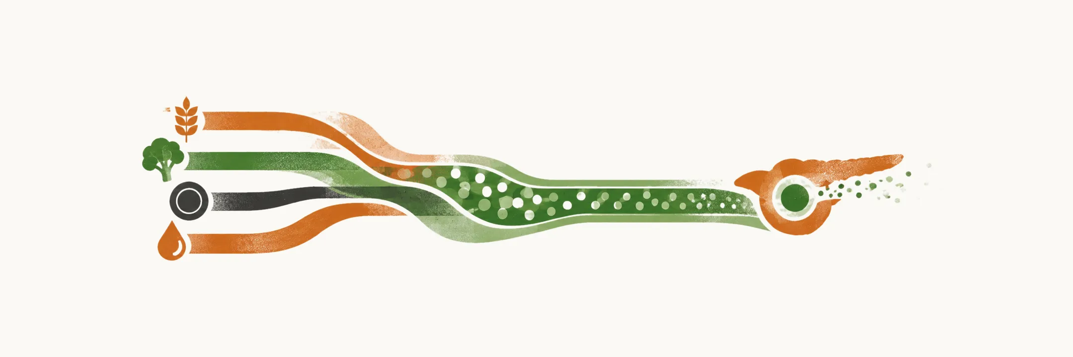
Blood glucose is held level by a balance between glucose coming in and glucose going out. Glucose comes in from the body’s own output and from the meal. It goes out into the tissues, mainly muscle, and the kidney takes back most of what it filters. After a meal, gut hormones add to the insulin response and hold back glucagon. Insulin then holds the liver back and pulls glucose into muscle.
In type 2 diabetes several parts of this balance respond less, and each weakened part pushes the others the same way. The eight mechanisms are the routes by which glucose is made too freely, taken up too poorly or kept too readily, while the pancreatic β cells fall behind.
Why does no single fault explain type 2 diabetes?
Several mechanisms act together to raise glucose in type 2 diabetes mellitus. No single one explains the disease, and eight are recognised. The earlier notes gave the balance. This one asks what can tilt it.
What does the gut add after a meal?
The first two mechanisms sit in the gut and the pancreas, so the first question is the gut’s contribution. After a meal the gut releases incretins. The gut releases these hormones as food arrives, most strongly with carbohydrate and fat, so the signal begins with the meal itself.
Incretins also reduce alpha cell activity and so lower the production of glucagon. GLP-1 holds glucagon down when glucose is above the fasting level, so after a meal the alpha cells are quieted and glucagon falls. This matters because glucagon is the hormone that drives hepatic glucose output. It tells the liver to break down its stored glycogen and to make new glucose, so it sets how much glucose the liver adds.
What goes wrong with the gut’s signal in type 2 diabetes?
The first mechanism is a decreased incretin effect. The gut’s added push on insulin after a meal is weaker in type 2 diabetes, and this loss is likely to contribute importantly to high glucose after meals.
The loss also reaches the alpha cells. In type 2 diabetes the gut’s signal is weaker, and GLP-1 secretion is itself impaired, so post-prandial inhibition of the alpha cells is reduced. Glucagon is then less restrained. The liver keeps releasing glucose after eating, because glucagon is what raises its output, and endogenous glucose production rises further.
What does the pancreas do when the gut’s help is less?
Insulin is the other half of the same balance. The second mechanism is decreased insulin secretion. In the early stages of the disease the β cells release more and more insulin. This compensation fails sooner or later, because insulin resistance keeps increasing. The β cells can raise their output for a time, but when resistance keeps rising they can no longer keep pace.
Where does insulin stop working in the body’s tissues?
The pancreas can only push so far, so the next question is what the tissues that insulin targets do. Insulin is the signal that holds the liver’s glucose output down and pulls glucose into muscle, as the normal glucose homeostasis note sets out. The third to sixth mechanisms are insulin-resistant tissues, including the kidney’s handling of glucose.
The third mechanism is insulin resistance of peripheral tissue, especially in skeletal muscle. Muscle is the main site of insulin-stimulated glucose uptake after a meal, so when it responds poorly, less glucose leaves the blood.
The fourth mechanism is increased hepatic glucose production. After a meal, insulin normally stops the liver adding more glucose. Here the liver is insulin-resistant and does not respond to that signal, so hepatic glucose production is not reduced after hyperglycemia. The liver keeps adding glucose when it should stop.
Can the brain change the liver’s output?
The liver is not the only organ with a link to insulin that matters here. The fifth mechanism is insulin resistance of the central nervous system, mainly the hypothalamus. When the brain’s insulin receptors, especially in the hypothalamus, stop responding, the liver makes more glucose.
The route is nervous. Hypothalamic insulin resistance raises sympathetic activation. Sympathetic activation increases endogenous glucose production.
The same central circuits are a target of incretins, mainly GLP-1. GLP-1 receptors are found in the hypothalamus. In experimental work, activating GLP-1 receptors in the brain reduced food intake, and GLP-1 acts on the central nervous system to decrease appetite.
What happens to glucose at the kidney?
The last place glucose is kept or lost is the kidney. The kidney takes back most of the glucose it filters. The sixth mechanism is enhanced glucose reabsorption by the tubular cells of the kidney. A higher take-back threshold has been proposed as a cause of high glucose, but this is not yet firmly supported. Glucose that would otherwise be excreted is retained, and hyperglycemia persists.
What does fat tissue add?
The last two mechanisms come from adipose tissue, which also sends fatty acids into the blood that the other tissues must handle.
The seventh mechanism is increased lipolysis. Insulin normally holds back the breakdown of fat in fat cells. The adipocytes lose this inhibitory effect, so lipolysis increases and free fatty acids reach the circulation.
The eighth mechanism is a chronic low-grade inflammatory state of adipose tissue. Inflamed adipose tissue releases cytokines, and these cytokines sustain the inflammatory state. Chronic low-grade inflammation has been suggested to raise the chance of insulin resistance, and it has been suggested to keep insulin resistance in place.
Why do the eight add up to one disease?
The mechanisms reinforce each other. Treatment commonly has to act on more than one mechanism, which is why they reinforce each other and treatment must target several.
Together the eight describe a disease in which glucose is produced too freely, taken up too poorly and reabsorbed too readily. The pancreatic β cells cannot make up the difference. But what starts the first tilt?