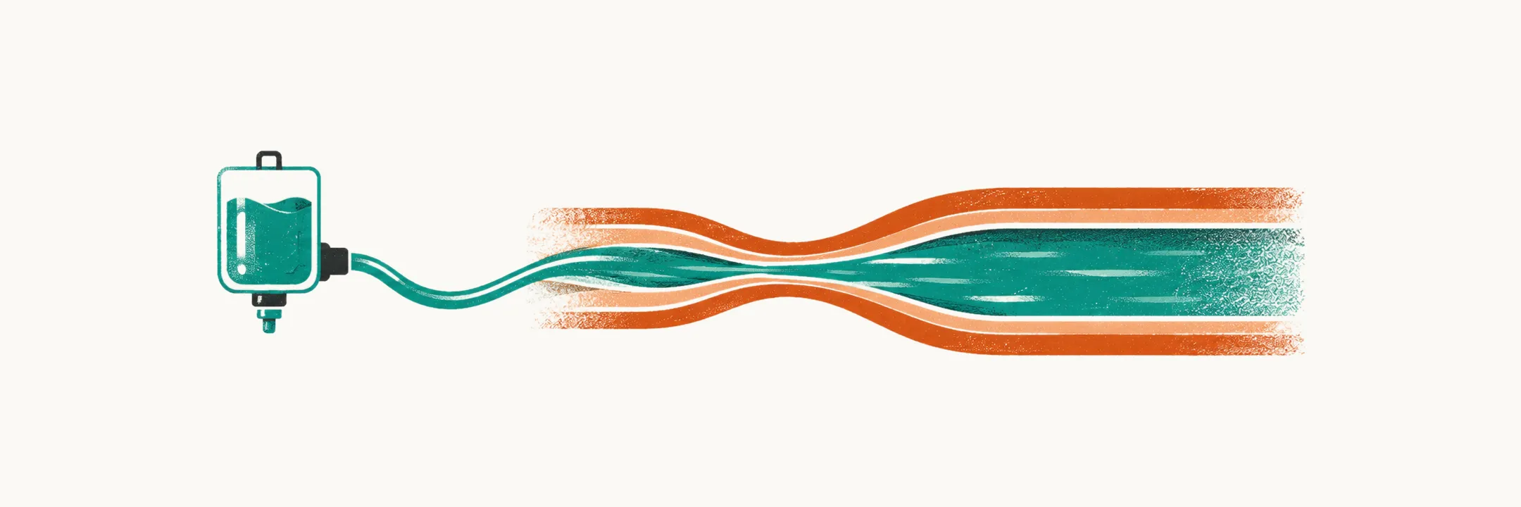
Most children with acute gastroenteritis are managed at home. Care steps up in stages: medical review, then hospital admission, then intravenous fluid. Each step has its own criteria.
Shock is the far end of this ladder. It is defined by tissues not receiving enough oxygen delivery. The first minutes of shock treatment follow a set protocol. Some of these children need to be seen, so the first question is which ones.
Which children need to be seen?
The first step is deciding when a child cared for at home needs to be seen. The first step up is a review by a clinician, and it has its own list.
For infants and toddlers, the 2014 ESPGHAN/ESPID guideline recommends referral for medical evaluation when any of the following is present:
- age under 2 months;
- severe underlying disease, such as diabetes or renal failure;
- persistent vomiting;
- high-output diarrhea, with more than 8 episodes a day;
- family-reported signs of severe dehydration.
The vomiting and stool criteria describe the usual ways a child loses fluid. Vomiting is a common cause of dehydration in children, so repeated vomiting is a fluid loss in its own right. Diarrhea is the other common cause, and more stools mean more water lost. Younger children reach dehydration sooner, as the earlier note showed.
How firm is that advice?
The list is firm in its wording but thin in its evidence, and some of its items are also risk factors. Each referral criterion is a strong recommendation, and each is made on low or very low quality evidence.
Several of these criteria also appear among the risk factors for severe dehydration: young age, frequent stools and repeated vomiting. They appear on both lists because they are the same losses, or the same ways of losing fluid.
When does a child need admission?
Admission is the next step up, and the guidance here is even less firm. Evidence is thinner at this step. Admission decisions are consensus-based. The guideline states that there are no formally established admission criteria for acute gastroenteritis, and case-controlled studies cannot be done for ethical reasons.
What does the admission list contain?
The list is consensus, so each item is a reason the child cannot safely stay at the lower step. It includes:
- shock;
- severe dehydration (more than 9% of body weight);
- neurological abnormalities such as lethargy or seizures;
- intractable or bilious (bile-stained) vomiting;
- failure of oral rehydration;
- a suspected surgical condition;
- conditions for safe follow-up and home management not met.
Why is safe follow-up the criterion most often missed?
Safe follow-up is the criterion most often missed. A child with a modest deficit whose caregivers cannot reliably give oral rehydration solution at home is not a safe discharge. Yet the guideline records social and logistical concerns as a questionable indication for admission. So this criterion is a judgement call rather than a rule.
Which route of rehydration comes first?
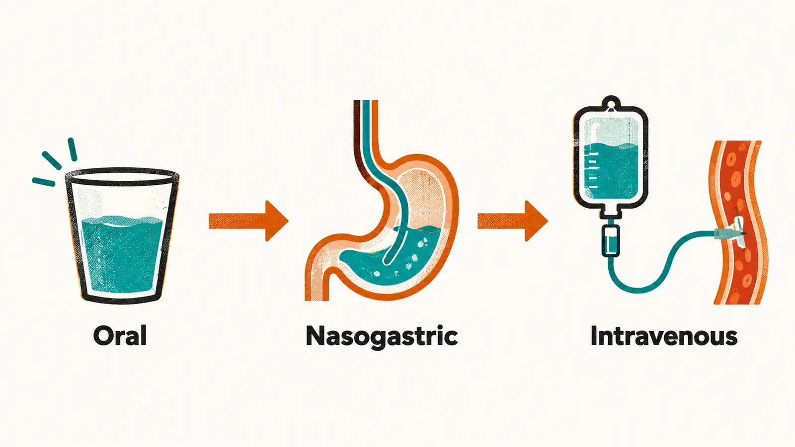
Whatever the admission decision, the fluid still has to go in, so the route is the next question. The route is chosen in steps. Oral rehydration is first-line. It works because the gut absorbs sodium together with glucose through a shared carrier, and oral rehydration solution is built on that cotransport. Water is thought to follow the sodium and glucose.
When oral intake is not feasible, enteral rehydration through a nasogastric tube should be proposed before intravenous rehydration.
What does the nasogastric route gain?
Before a vein is used, the nasogastric route is proposed. Compared with intravenous rehydration, it has significantly fewer major adverse events and a shorter hospital stay. It succeeds in most children. Rapid nasogastric rehydration, 40 to 50 mL/kg over 3 to 6 hours, is as effective as the standard 24-hour regimen.
When does rehydration move to a vein?
The move to intravenous fluid has its own consensus-based list, and its first item is shock. Intravenous escalation is indicated for:
- shock;
- dehydration with altered level of consciousness, a sign that perfusion is poor, because the brain is one of the organs whose perfusion falls when pressure falls;
- severe acidosis, because diarrhea can lower bicarbonate directly, as bicarbonate is lost through the gut;
- worsening dehydration, or lack of improvement despite oral or enteral rehydration;
- persistent vomiting despite appropriate fluid administration orally or via nasogastric tube;
- severe abdominal distension and ileus, in which the bowel stops propelling its contents.
What is shock?
Shock is the far end of the ladder, so the next question is what it looks like. It is the point at which intravascular volume has fallen far enough that tissue perfusion fails. Dehydration from diarrhea primarily comes from the intravascular volume shrinking while the intracellular volume is kept, and dehydration takes plasma volume down with it, as the earlier note showed.
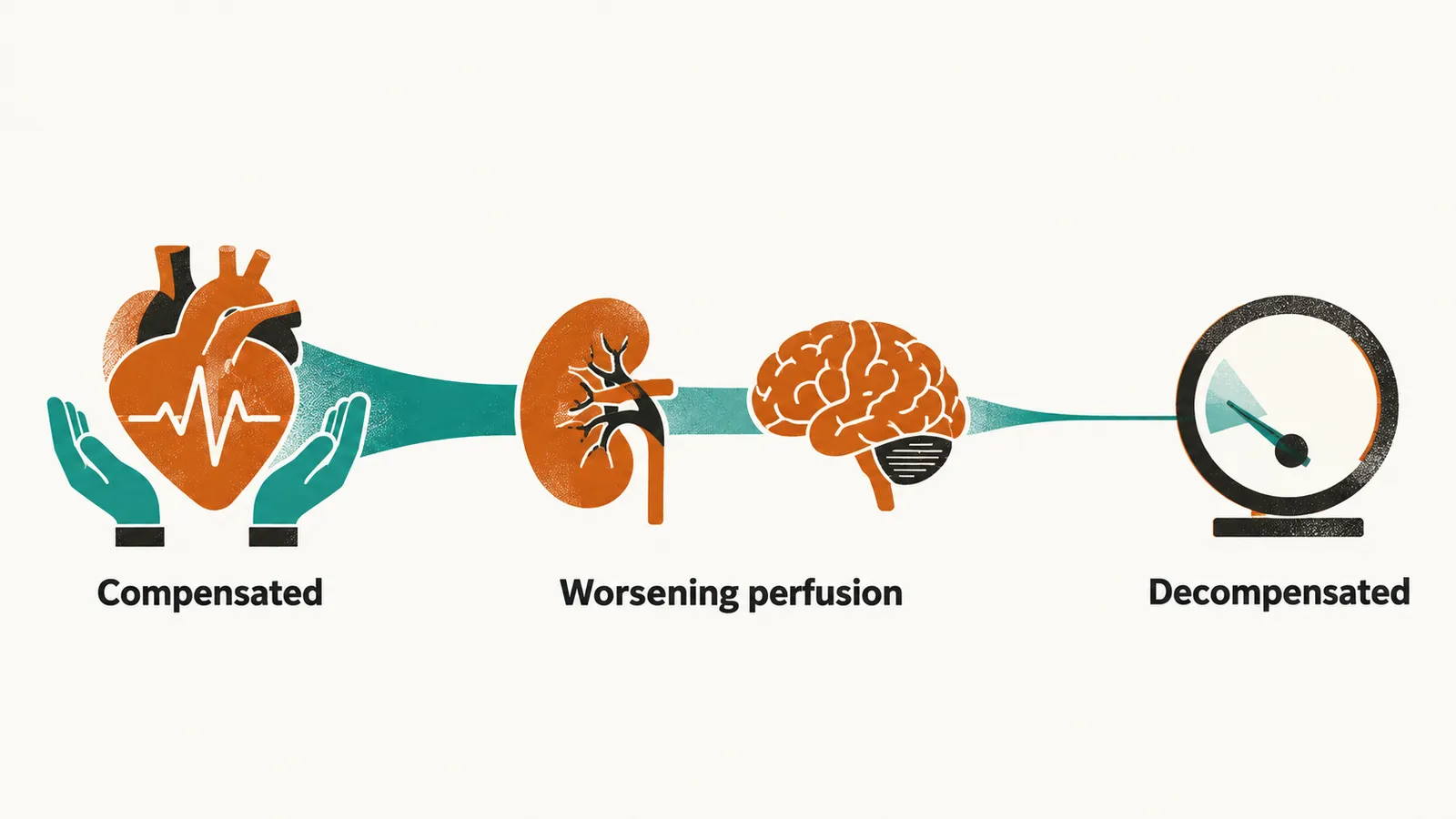
Shock is recognised in stages, because the body first compensates through compensatory responses.
What does compensated shock look like?
Early shock looks like a body that is still coping. When blood pressure starts to drop, the body triggers a rise in heart rate and peripheral vasoconstriction, and these hold the pressure up. The child is tachycardic with cool extremities, because vasoconstriction narrows the vessels of the skin. The child may look restless or agitated. Blood pressure is still normal, because the compensation is what keeps it so.
What shows that perfusion is getting worse?
Once the coping starts to fail, the organs show it. When systemic pressure drops, organ perfusion is impaired. The kidney is one of those organs, so urine output falls. Tachypnea appears. The brain is another, so the level of consciousness changes. Vasoconstriction diverts blood toward the core, so the periphery stays cold. The periphery also stays mottled.
Why is low blood pressure a late sign?
In decompensated shock the compensation can no longer hold, so blood pressure falls and organ perfusion is impaired. Hypotension is a late sign in most types of pediatric shock, because the compensatory responses hold the pressure up while they work. For the same reason, normal blood pressure does not exclude shock.
Why can the heart rate mislead?
Heart rate is one sign that compensation is working, and it can mislead in both directions. A slow heart rate in a dehydrated, poorly perfused child is a peri-arrest finding rather than a reassuring one. Pediatric resuscitation algorithms treat bradycardia with poor perfusion as impending arrest.
In the other direction, tachycardia in a child with diarrhea is not specific. Fever, pain and agitation all produce tachycardia. So tachycardia is read together with perfusion rather than on its own.
How is shock treated first?
Treatment starts with a bolus of fluid aimed at the vessels. Children presenting with shock secondary to acute gastroenteritis should receive a rapid intravenous infusion of an isotonic crystalloid as a 20 mL/kg bolus. The fluids named are 0.9% saline or lactated Ringer’s solution, both salt solutions in water. The aim is intravascular volume expansion, and both fluids serve it, because isotonic fluid primarily expands the extracellular compartment.
Why must the fluid be isotonic?
The bolus has to stay in the vessels, and that depends on the tonicity of the fluid. An isotonic fluid matches the concentration of plasma and primarily expands the extracellular compartment, so it stays out of cells. The bolus must be isotonic, not glucose-containing.
Why must the bolus be rapid?
The bolus must be genuinely rapid; the stated window is 10 to 15 minutes. Preload is the filling of the heart that the bolus is meant to restore, and a slow infusion does not achieve the same preload effect.
What if blood pressure does not improve?
If the first bolus does not help, the plan changes, and fluid loss may not be the whole cause. If blood pressure has not improved after the first bolus, a second, or even a third, bolus of 20 mL/kg should be given over 10 to 15 minutes. Other possible causes of shock should also be considered. The shock bolus recommendation is strong, on very low quality evidence.
Beyond three boluses, reconsider the diagnosis. In a child with diarrhea and vomiting the cause is usually hypovolemic, because the loss falls on intravascular volume. Failure to respond should prompt a search for septic, cardiogenic, anaphylactic, adrenal or obstructive causes.
What does the bolus not do?
The bolus is the first part of the treatment. It is resuscitation, not restoration, because it refills the intravascular part of the loss while the intracellular volume is kept. It does not replace the full fluid deficit. It does not replace ongoing stool and vomit losses. It does not correct electrolyte or acid-base abnormalities, and it does not treat the underlying infection.
Those aspects are handled in the phases that follow, set out in Rehydration Management in Acute Diarrhea. The severity grade and sodium pattern that decide those phases come from Clinical Assessment of Acute Diarrhea and Dehydration. Once the circulation is steadied, what replaces the rest of the deficit, and when?