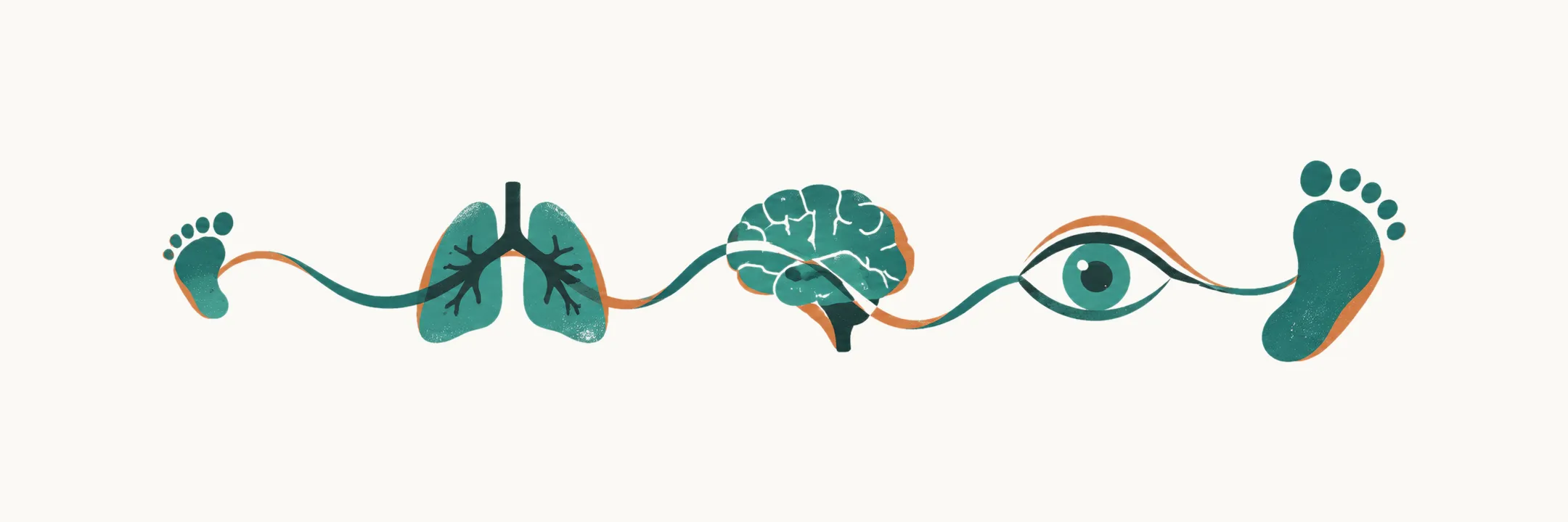
Survival does not end the disease of prematurity. Preterm means born before 37 completed weeks, as the corrected-age note set out. An infant born that early may move from acute respiratory and circulatory failure into weeks or months of lung support, feeding dependence, infection risk, sensory injury and neurodevelopmental vulnerability. So the child leaves intensive care with several injuries to follow, and the first question is which of them come first.
When does each risk arrive?
The timeline of mortality and morbidity follows the order in which immature organs meet the demands of life outside the womb. Each interval therefore has its own substrate.
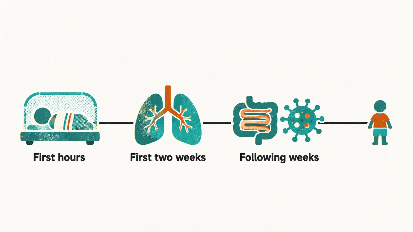
In the first hours, profound whole-body immaturity may be incompatible with life outside the uterus. As the golden-hour note explained, the first hour is when the placenta’s support stops and the infant must do the job alone. Even when the body is mature enough, the transition itself can fail, and as the golden-hour note explained, the change from fetal to newborn circulation and breathing can fail.
In the first two weeks, respiratory distress syndrome, lung failure caused by surfactant deficiency, becomes a major cause of death. As the respiratory note explained, a shortage of surfactant lets the small airspaces collapse early. Circulatory transition and early brain injury also become major causes of death. As the ductus note and the brain-injury note explained, the circulation and the brain can each fail early.
In the following weeks, three things dominate:
- Necrotizing enterocolitis, inflammatory necrosis of immature bowel. As the nutrition note explained, the immature bowel wall is injured through several contributors, not one cause.
- Hospital-acquired infection. As the infection note explained, the infant’s defences are borrowed and immature, and lines and devices give organisms a route in.
- The consequences of prolonged intensive care. The NICU environment, and the lines and devices it uses, add their own load, as the golden-hour and infection notes explained.
So the answer to “what is this infant at risk of now?” differs between week one and month three.
What carries forward out of the admission?
Most morbidity that matters later is established during the neonatal weeks and expresses itself over years. The lung shows why. A lung injured while it is still growing cannot catch up, so the limit the injury sets persists, even into adulthood.
That is why chronic lung disease can remain a life-threatening respiratory condition beyond the acute admission. The chronic lung disease here is the bronchopulmonary dysplasia the respiratory note described: a lung that is still growing and has been injured. Neurodevelopmental, visual and growth problems, by contrast, become apparent over months and years.
One child can be followed for lung, gut, brain and eye consequences at the same time. Two of these, brain injury and retinopathy of prematurity, decide much of the long-term picture.
Why is the lung the most visible legacy?
Bronchopulmonary dysplasia (BPD) is also called chronic lung disease of prematurity. It is the best-characterized chronic complication after extremely preterm birth. As the respiratory note described, it develops in a lung still trying to grow while exposed to oxygen, inflammation, ventilator stress and fluid overload.
Postmenstrual age is gestational age plus the completed weeks since birth, as the corrected-age note set out. Among extremely preterm infants surviving to 36 weeks postmenstrual age, about half meet an oxygen-use definition.
That phrase, an oxygen-use definition, matters because definitions of BPD differ. As the respiratory note explained, BPD is defined in more than one way, and the definition decides who is counted. The incidence therefore depends on which definition is used.
What does the lung need after discharge?
Once the diagnosis is made, the next question is what the child needs at home. Affected infants may stay on respiratory support and saturation monitoring for weeks or months. In severe cases infants go home with oxygen. Rarely, severe cases go home with tracheostomy and long-term ventilatory support.
The chest imaging follows the same range of severity. It runs from fine granular opacity in moderate disease to coarse interstitial change, hyperinflation, fibrosis or persistent ventilation in severe disease.
The lung is the most visible legacy, but not the only one.
Why does feeding lag behind?
Affected infants may feed late because breathing consumes their energy budget. As the respiratory note explained, each breath costs energy, so a tiring infant has less left for feeding.
Feeding autonomy is often the last milestone of the neonatal admission. Respiratory support, immature suck–swallow coordination and increased work of breathing in BPD all interfere with oral feeding. Sucking, swallowing and breathing have to be timed together, and that coordination is an immature system that matures late, as the corrected-age note explains.
The gut adds its own limit. Bowel resection after necrotizing enterocolitis can reduce absorptive surface. The gut absorbs through its inner surface, so when a length of bowel is removed that surface goes, and what remains absorbs less and moves food faster.
What does the brain carry forward?
Brain injury is the other legacy that decides much of the outcome, and its frequency falls as gestational age rises. As the brain-injury note explained, the germinal matrix shrinks as the brain matures, so bleeding from it becomes less likely.
The injury takes several forms:
- Germinal matrix hemorrhage: bleeding from a fragile vascular region beside the ventricles. The bleeding from the germinal matrix is the one the brain-injury note described.
- Periventricular hemorrhagic infarction: usually a venous infarction of brain tissue beside the ventricle. As the brain-injury note explained, the fragile vessels beside the ventricles can rupture.
- Cystic periventricular leukomalacia: small cysts left by injury to the periventricular white matter. As the brain-injury note explained, the white matter beside the ventricles can be injured without any bleed, because its oligodendrocytes are vulnerable.
Brain injury is the substrate of most later cerebral palsy among preterm survivors. Most immature survivors carry the highest risk of spastic motor impairment. They also carry a higher risk of cognitive, language and behavioral difficulty. Outcome here is multifactorial, and it is thought to reflect an immature brain together with perinatal and environmental factors. Cognitive, language and behavioral difficulty does not show itself in the same obvious way as a motor impairment.
What does the eye carry forward?
The eye is the other organ that decides much of the long-term picture. Retinopathy of prematurity is abnormal growth of retinal vessels that had not finished developing at birth. As the retinopathy note explained, the retinal vessels were still unfinished at birth and grow abnormally when injured.
Even when the acute disease has resolved, it may leave myopia, strabismus or refractive error. For this reason the eyes still need review after discharge.
Retinopathy of prematurity remains a leading cause of childhood blindness where screening and treatment are not available. As the retinopathy note explained, screening is timed to vessel growth, so where screening is absent the disease is missed. Treatment of severe retinopathy of prematurity markedly reduces that outcome. As the retinopathy note explained, treatment acts on the starved retina or on its growth signal.
How is growth read?
Growth cannot be interpreted without the lung, gut and neurological history.
Growth and development are interpreted using corrected age. As the corrected-age note explained, corrected age counts from the due date, not from birth. As the corrected-age note explained, development is read against corrected age rather than chronological age alone. A child born 12 weeks early reaches some milestones later by chronological age even when development is appropriate for corrected age, as the corrected-age note explained. For growth in extremely and very preterm children, age correction may be needed out to 36 months.
Who follows the child, and how?
Follow-up has two jobs: to track the organ injuries accumulated during intensive care, and to interpret later growth and development using corrected age. Follow-up programmes monitor domains that can fail silently. These are:
- respiratory symptoms and lung function where relevant
- growth
- feeding and nutrition
- hearing
- vision
- motor development
- language and cognition
Exactly which tests are done, and at what intervals, follows local programme protocol. Published schedules differ, and thresholds change with evolving evidence.
Who watches between the visits?
The family is part of monitoring, and the schedule is left to the local programme, so the question is who watches between visits. A prolonged admission changes family life. Parents may be separated from work, home and other children for months. They were often prepared for a healthy term newborn rather than an infant on respiratory support. Family-centered care keeps parents involved during the admission.
After discharge, parents observe breathing, feeding, growth and development every day. Their reports are clinical information. So when something changes between visits, who is most likely to notice it first?