
The anterior pituitary is built from five kinds of hormone-producing cell. A pituitary adenoma is a benign tumour derived from one of these five. Because the tumour keeps making the hormone of the line it came from, the cell type sets the hormonal picture. So the useful way to sort adenomas is by the cell they came from. Which line gives which tumour?
Which cell gives which adenoma?
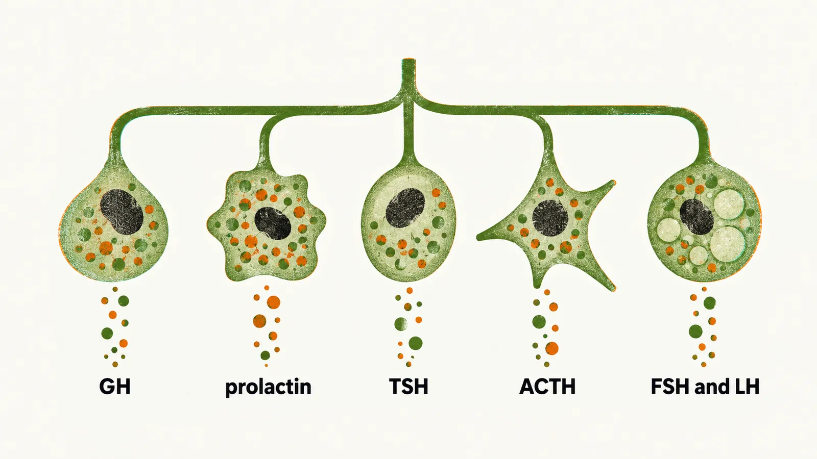
If every adenoma comes from one of five lines, the next question is which line gives which tumour and which hormone. Each hormone-producing cell lineage gives its own adenoma, so the cell of origin is the natural way to sort them.
- Somatotroph adenomas produce GH. They make up 10-15% of pituitary adenomas.
- Lactotroph adenomas produce prolactin. They are the most common form of functioning pituitary adenoma, and their prevalence is 50-60% of all functioning pituitary adenomas.
- Thyrotroph adenomas produce TSH.
- Corticotroph adenomas produce ACTH.
- Gonadotroph adenomas produce FSH and LH.
What does an adenoma do to hormone levels?
Each line makes its own hormone, so the next question is what the tumour does with that hormone. Pituitary adenomas are the most common cause of pituitary hypersecretion syndrome, and also of hyposecretion in adults.
Hormonally active pituitary adenomas work autonomously. Feedback has a reduced effect on them. A normal cell responds to circulating hormone levels and throttles its output. The tumour does not, so its cells keep secreting when the feedback signal says stop, and hormone output can rise above normal.
One more point concerns size. The size of a pituitary adenoma has no correlation with the level of hormone secretion from the tumour. The study behind this looked at ACTH-secreting adenomas, so it should not be stretched further than that.
Can something outside the pituitary drive the same cells?
Hormone output has one more source to consider, a signal from outside the gland. GHRH and CRH are able to cause hyperplasia of their target cells in the anterior pituitary. And in some rare tumours of the abdomen and chest that produce GHRH and CRH, hyperplasia of somatotroph and corticotroph cells in the pituitary is possible.
Which adenomas make no hormone, or more than one?
Not every adenoma makes a hormone of its own, and some make more than one. The non-functioning pituitary adenoma is a sixth clinical group. It does not produce a syndrome from hormone excess. It accounts for 15-35% of all cases.
Besides the five cell types, polyhormonal adenomas are possible. They are either mixtures of different cell types, or one cell type able to produce different hormones at the same time.
How are adenomas sorted in the lab?
To name the line behind a tumour, pathologists use the markers each line depends on. Within each lineage, adenomas are further typed by the transcription factors they express, the hormones they contain and their cytokeratin staining pattern. Pathologists stain for the three main transcription factors so that the line can be named.
- Pit-1 marks GH-, PRL- and TSH-producing adenomas. Pit-1 is needed for the development of the somatotroph, lactotroph and thyrotroph lines, which is why these three share one marker.
- Tpit marks ACTH-producing adenomas.
- SF-1 marks gonadotroph adenoma.
The profiles that follow use the abbreviations GH, PRL, α-SU and ER.
What do the stains show for the other types?
The markers for the other lines give a clear pattern. Each profile lists transcription factors, then hormones, then the cytokeratin pattern.
Lactotroph line:
- Sparsely granulated lactotroph adenoma: Pit-1, ER; PRL (Golgi); cytokeratin diffuse.
- Densely granulated lactotroph adenoma: Pit-1, ER; PRL (diffuse); cytokeratin diffuse.
- Acidophil stem-cell adenoma: Pit-1, ER; PRL (diffuse), GH; cytokeratin rare and dot-like.
Thyrotroph line:
- Thyrotroph adenoma: Pit-1, GATA-2; β-TSH, α-SU; cytokeratin diffuse.
Corticotroph line:
- Densely granulated corticotroph adenoma: Tpit; ACTH; cytokeratin diffuse.
- Sparsely granulated corticotroph adenoma: Tpit; ACTH; cytokeratin diffuse.
- Crooke’s cell adenoma: Tpit; ACTH; cytokeratin ring-like.
Gonadotroph line:
- Gonadotroph adenoma: SF-1, GATA-2, ER; β-FSH, β-LH, α-SU; cytokeratin diffuse.
Adenomas that fit no single line:
- Silent type III adenoma (plurihormonal): Pit-1 (?), ER; multiple hormones; cytokeratin diffuse.
- Unusual plurihormonal adenoma, not otherwise specified: multiple transcription factors; multiple hormones; cytokeratin not applicable.
- Null cell adenoma (hormone negative): no transcription factors, no hormones; cytokeratin diffuse.
Why are there two kinds of somatotroph adenoma?
The somatotroph line splits into two kinds under the microscope. Somatotroph adenomas are divided into two variants. They differ in how many GH-containing granules the cells hold. Densely granulated somatotroph adenomas (DGSA) are the more common form, and their cells look like normal somatotrophs because they hold many granules. Sparsely granulated somatotroph adenomas (SGSA) are less common, and their cells hold only a few granules.
SGSA are more prevalent in younger adults and in females, and they have more potential for invasion. They also express less growth hormone positivity than DGSAs. Mixed-pattern somatotroph adenomas, containing both variants, are also possible.
The profiles are:
- Densely granulated somatotroph adenoma: Pit-1; GH, α-SU; cytokeratin diffuse.
- Sparsely granulated somatotroph adenoma: Pit-1; GH; cytokeratin dot-like.
- Mammosomatotroph adenoma: Pit-1, ER; GH, PRL, α-SU; cytokeratin diffuse.
- Mixed somatotroph and lactotroph adenoma: Pit-1, ER; GH, PRL, α-SU; cytokeratin diffuse.
Can a pituitary adenoma become a carcinoma?
Adenomas are benign, so the next question is when one counts as malignant. Pituitary carcinoma is very rare. It is defined by spread, not by how the tumour looks: a pituitary tumour that has spread beyond the sella to the cranium, the spine or the rest of the body. Without spread, nothing in the histology separates it from an aggressive adenoma.
A malignant tumour involving the pituitary is far more often a metastasis that travelled from elsewhere, since pituitary metastases come from cancers outside the gland.
How common are pituitary adenomas?
Carcinoma is very rare, so the common case is the benign adenoma. How common is it? The prevalence of pituitary adenomas is about 1:1000-1500. Yet autopsy studies find that about 15% of individuals have completely silent, asymptomatic pituitary adenomas, and a pituitary microadenoma is found in about 1/4 of all autopsies. Pituitary adenomas are more common in women during fertile ages, and they account for about 15% of all intracranial neoplasm cases.
What makes one of these cells start to grow into a tumour in the first place?