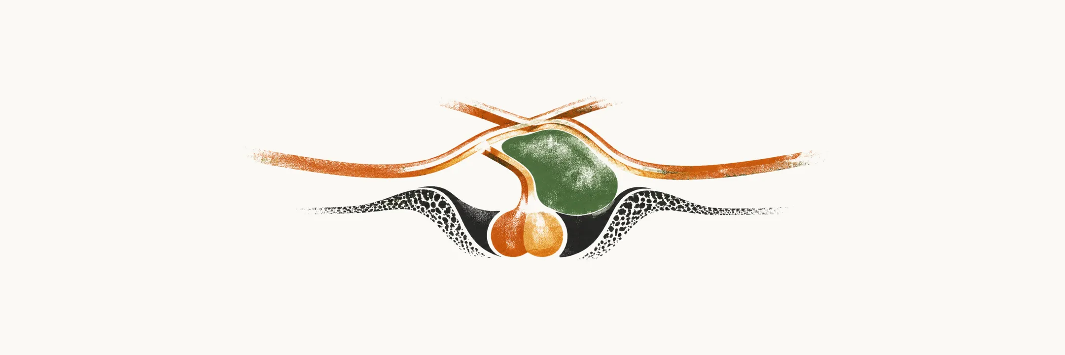
The sella is the bony cup that holds the gland. It has bone on every side and a tight membrane as its roof, so a mass that grows inside it cannot simply widen the space. Next to it lie the optic chiasm above, the cavernous sinuses on each side, and the hypothalamus above the gland. A growing mass can therefore reach all three.
The pressure falls first on the nearest structure. So the picture is simple: what a sellar mass does to a patient follows from where it pushes.
Why is headache so often the first sign?
Because the sella has no room to widen, a mass that fills it raises the pressure inside. Headaches are a common feature of pituitary tumours, even when the tumour is very small. They also occur without any suprasellar expansion.
Why should a small mass hurt? The gland is confined, so small changes in pressure stretch the dura mater, and that stretching causes headache. This mechanism is proposed rather than settled. It may also explain why headache can occur when the mass does not bulge upward at all.
Which way does the mass grow, and does size matter?
Pressure inside the sella has to go somewhere, so the next question is which way the mass goes. The effect on the patient depends on three things: the anatomic location of the mass, the direction in which it extends, and its size. A larger mass presses harder on what is next to it.
The most frequent direction of expansion of pituitary adenomas is suprasellar, upward out of the sella. A soft mass usually indents the diaphragm sellae, which is why upward growth is the usual route.
Can the mass grow through the bone?
Upward growth meets the diaphragm, and downward growth meets bone. Adenomas are benign in their cells but can grow into the tissue next to them, and bony invasion by pituitary tumours is possible. It happens mostly in the floor of the sella turcica. The sphenoid sinus lies directly below the sella, so erosion of the floor leads to invasion of that sinus.
Aggressive tumours can, rarely, invade the roof of the palate. When they do, they can cause nasopharyngeal obstruction, infections and CSF leakage. The leak is possible because, once the bone is open, the CSF space around the sella connects with the air-filled sinus, and fluid can come out through the nose.
Why does vision fail?
Upward growth is the usual route, and that route reaches the optic nerves. Vision loss from a mass above the gland can come from pressure on the optic pathway, and suprasellar expansion can cause visual disturbances in several different ways.
The most common mechanism is compression of the optic chiasm, the point where the two optic nerves meet. The chiasm sits just above the gland, so upward growth reaches it first. The other ways are these:
- The tumour can invade the optic nerves directly.
- The tumour can compromise CSF flow, with a secondary effect on the visual system through hydrocephalus.
Why do the eye movements fail?
A mass that grows to the side meets a different neighbour, the cavernous sinus. The cavernous sinuses lie on each side of the gland, and their lateral wall carries cranial nerves III, IV, V1, V2 and VI. Expansion to the side can therefore damage the nerves that run beside the gland.
Depending on the extent of the neural damage, patients can have neurological manifestations due to invasion of cranial nerves III, IV and VI. These nerves move the eyes, so when they are hit the eyes stop moving together. Cranial nerve involvement can lead to diplopia, ptosis, strabismus and ophthalmoplegia. V1 and V2 run in the same wall, so loss of facial sensation can also occur.
What happens to the stalk when it is pressed?
The cavernous sinus is at the side; the stalk runs down the middle to the gland. The stalk carries the hypothalamic signals down a portal blood supply to the front of the gland. Compression of the stalk can decrease portal system flow, and so it can decrease the passage of neurohormones and dopamine from the hypothalamus to the pituitary gland.
Dopamine from the hypothalamus is a brake on prolactin, so cutting its flow lets prolactin rise. That is why stalk compression results in early hyperprolactinemia.
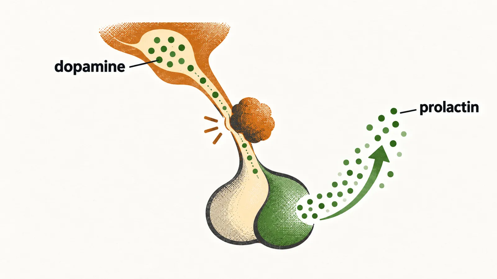
The other hormone-making cells of the front gland also depend on signals from above. As the stalk stays pressed, they lose that support, and stalk compression later results in concurrent loss of the other pituitary hormones. A formed pituitary tumour causes harm in two ways: through the hormones it secretes and through the structures it compresses. The stalk is a clear example of the second.
What does the hypothalamus control, and what is lost when a mass reaches it?
The stalk is the link to the hypothalamus, so the next question is what the hypothalamus itself does. A lesion that reaches the hypothalamus can bring hypothalamic problems with it, because the hypothalamus runs many functions. Pituitary tumours can therefore cause metabolic alterations. They include:
- precocious puberty
- hypogonadism
- diabetes insipidus
- sleep disturbance
- dysthermia (disturbed temperature regulation)
- appetite disorder
Some of these are easy to place. Hypothalamic tumours can over-produce GnRH. GnRH raises FSH and LH, so damage that cuts this drive leaves the gonads under-stimulated, which gives hypogonadism. ADH is made in the hypothalamus and stored in the posterior pituitary, so damage to the stalk or back lobe removes ADH, which causes diabetes insipidus.
Why do sleep, appetite and temperature go wrong?
The hypothalamus runs sleep and circadian rhythm through networks of activating and inhibiting neurons, so damage there disturbs sleep. It also controls appetite and weight, which explains appetite disorder. Temperature control sits there too, with the anterior part losing heat and the posterior part keeping heat. Damage disturbs that control, and the result is dysthermia.
Where in the hypothalamus is the damage?
The anterior and posterior regions have different jobs, so the pattern of signs can point to the location of hypothalamic involvement.
- Lesions involving the anterior and preoptic regions cause paradoxical vasoconstriction, hyperthermia and tachycardia. The preoptic area starts heat loss, so when it is damaged the body cannot shed heat and runs hot.
- Posterior hypothalamic involvement causes central disorders of thermoregulation. Signals from the front area are passed back to the posterior hypothalamus, so damage there disturbs the central control of temperature.
- Damage to the preoptic regions can cause polydipsia or hypodipsia, because thirst is one of the hypothalamic controls.
- Lesions of the central hypothalamus cause over-activation of the sympathetic system. The hypothalamus is the top of autonomic control, so a lesion can change sympathetic output. The over-activation is the clinical pattern; its mechanism is not settled.
What does sympathetic over-activity do?
Sympathetic over-drive causes arrhythmia, hypertension and gastric erosion. It speeds the heart and raises blood pressure, which explains the arrhythmia and the hypertension.
Why does apoplexy come on suddenly?
A mass that has grown slowly can also change in a short time. Pituitary apoplexy differs in being sudden. It is sudden bleeding or infarction into a pituitary tumour, which happens inside an existing adenoma, and its effects are sudden too. It complicates roughly 2-12% of pituitary tumours.
It most often occurs in a non-functioning macroadenoma. An adenoma that makes no hormone gives no hormonal warning, so it may be large when found. A proposed explanation is that the raised pressure inside such a tumour may cut its blood supply and cause ischaemic necrosis.
What does apoplexy look like?
Sudden bleeding causes the pressure picture above, all at once. Pituitary apoplexy presents with sudden onset of severe headache, vomiting, visual loss and meningism. The sudden swelling stretches the sella, so the headache is sudden, and the visual loss comes from pressure on the chiasm and nerves. Apoplexy can also reduce the level of consciousness.
Why is apoplexy an endocrine emergency?
The loss of the gland’s output is the next point. A mass can squeeze the normal gland, leaving it under-active, but a bleed or infarct destroys hormone-making tissue in a short time. Rapid expansion and necrosis leave most patients with acute hypopituitarism, so output stops at once rather than fading. That is why pituitary apoplexy is an endocrine emergency.
Loss of ACTH is the life-threatening part. ACTH drives cortisol, and cortisol is needed to hold blood pressure and glucose under stress.
What is done first?
Because ACTH loss is the danger, the first step is to replace what the gland can no longer make. Treatment begins immediately with glucocorticoid replacement and correction of fluid and electrolyte balance.
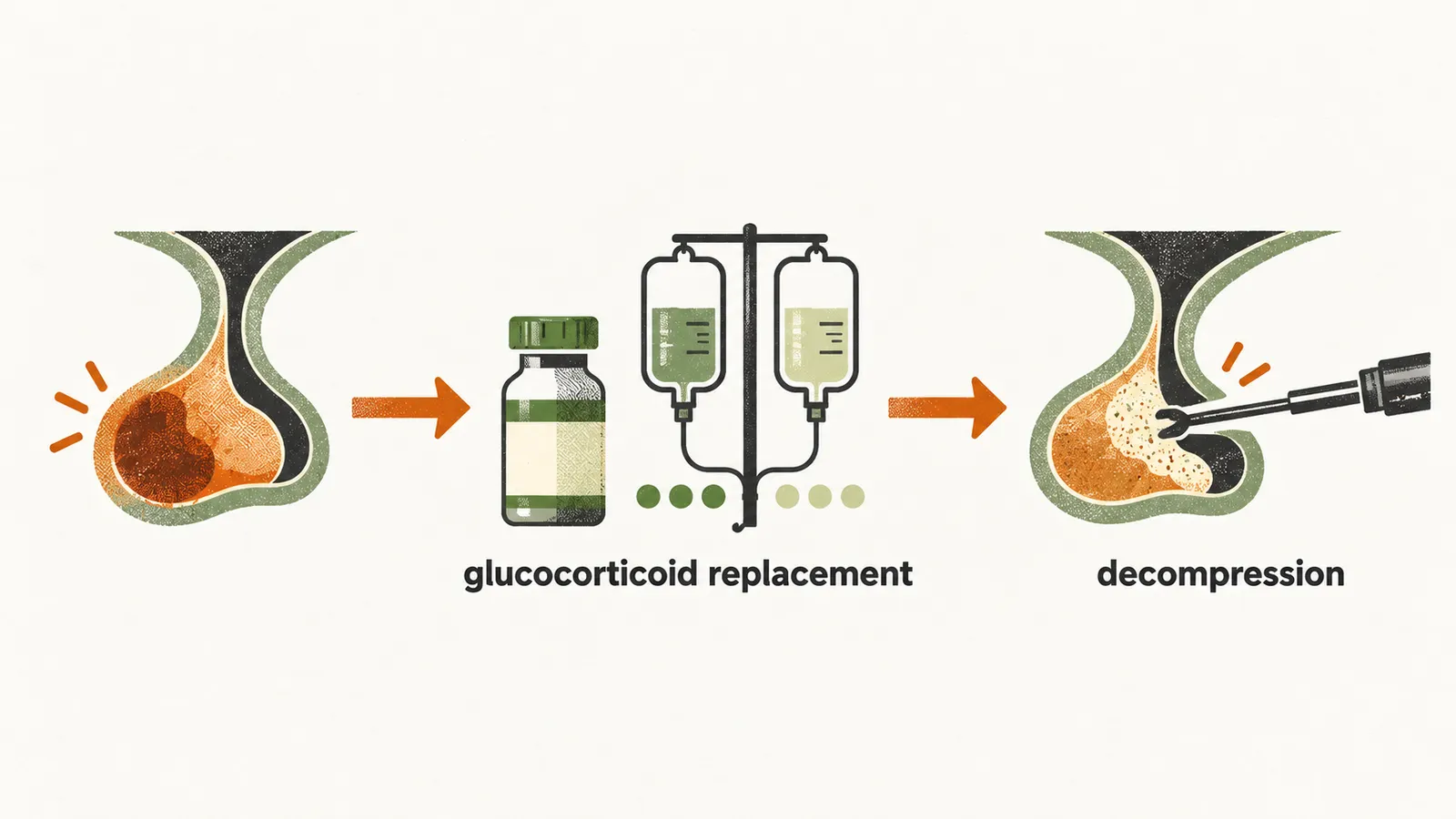
Transsphenoidal decompression is considered when visual acuity, visual fields or consciousness deteriorate. Removing the pressing tissue relieves pressure on the chiasm and nerves.
But how is a mass found in the first place, when headache or a visual complaint can come from many other causes?