
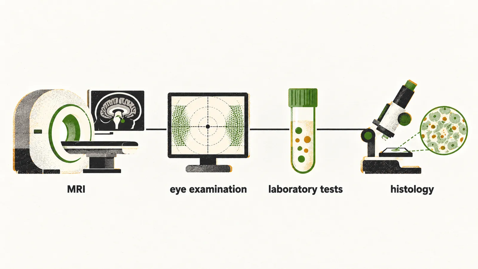
A mass in the sella, the bony cup that holds the pituitary gland, is investigated four ways: MRI, ophthalmologic evaluation, laboratory investigation and histologic investigation. Each answers its own question. MRI shows the mass and its surroundings. The eye examination tests the vision the mass threatens. The laboratory tests look for hormone excess or loss. Histology examines tissue when the other results do not settle the diagnosis. No test replaces another, and together they decide what happens next.
Why do most masses turn up on a scan?
The work-up often starts from a scan that was done for another reason. Sellar masses are mostly incidental findings on MRI, and most of them are pituitary adenomas. A mass discovered by chance in a scan done for another reason is called an incidentaloma.
What does MRI show, and how is it done?
The scan comes first because it shows where the mass is and what it touches. MRI separates soft tissues from one another. The gland, its stalk, the optic chiasm and the nearby nerves are all soft tissue, so MRI is the view that shows the mass against them.
The technique uses a contrast agent, gadolinium. Sagittal and coronal T1-weighted images are taken before and after gadolinium. This gives a precise picture of the hypothalamus, pituitary stalk, pituitary tissue, cavernous sinus, sphenoid sinus and optic chiasm.
Contrast helps the picture in more than one way. The normal anterior pituitary gland is thought of as heterogeneous tissue on MRI, because contrast reaches it in stages, starting at the stalk and spreading to the front of the gland, so the signal is not the same everywhere at every moment. An adenoma usually differs in density from the surrounding tissue, because it takes up gadolinium less well than normal gland.
Why is CT still used?
MRI is the main view, but CT has a role for bone and calcium. A CT scan is reserved for investigating the level of bone erosion and the calcification of the mass.
What does the eye examination test?
A scan can outline the mass, but it does not show whether the nerves beside the gland still work. The optic chiasm lies next to the gland, so a mass that grows upward can press on it. The pattern of vision loss depends on how the lesion sits against the chiasm. The eye examination therefore shows the effect of a mass that imaging only outlines.
Why do the outer halves of vision go first?
The classical ophthalmological finding in pituitary tumours has a particular pattern, and it follows from where the nerves cross. It is bitemporal hemianopia, partial blindness of the outer vision. It is more pronounced for superior vision.
The nasal fibres of each retina cross at the chiasm. When the crossing point is pressed, their loss takes out the outer field of vision on both sides. Nasal ganglion cell fibres are more vulnerable to compression of the ventral optic chiasm, and why they suffer first is thought to be explained by this vulnerability rather than being fully settled.
Why can a mass cause double vision?
When a mass spreads sideways, it is not the field of vision that fails but the movement of the eyes. The eye-moving nerves III, IV and VI run beside the gland, through the cavernous sinus, so a mass that spreads sideways can squeeze them. Tumour invasion into the cavernous sinus can therefore produce diplopia. It results from invasion of the ocular motor nerves located in the cavernous sinus, because a palsy of one of these nerves leaves the eyes unable to move together.
What do the hormone tests measure?
The second question is whether the mass has changed the hormones. A mass in the sella can leave the normal gland underactive or overactive, and adenomas can secrete their hormone in excess, which is the hypersecretion these tests look for. The laboratory investigation looks for hormone excess or loss.
The functional pituitary adenomas are Cushing’s disease, prolactinoma and acromegaly. In these, the clinical manifestation should guide the laboratory investigation. When a pituitary tumour is suspected on MRI, several tests are usually performed:
- Prolactin (PRL).
- IGF-1 (insulin-like growth factor 1).
- 24-hour urinary free cortisol.
- FSH and LH.
- Thyroid function tests.
Why is IGF-1 used to check growth hormone?
Growth hormone is not judged by measuring it in the blood directly. The liver makes IGF-1 in response to growth hormone. IGF-1 does not swing with sleep, exercise or the time of day the way growth hormone does, so it is a steadier measure of how much growth hormone the gland makes.
Why is cortisol measured in a 24-hour urine sample?
Cortisol needs a different sample, collected over a day. Part of circulating cortisol is free, and the free part is what the kidney filters into urine. Urine collected over 24 hours therefore reflects the cortisol the body released over that day, and that output correlates well with the rate of cortisol secretion.
When is a tissue sample needed?
When the hormone results do not settle the question, the tissue itself is examined. Histological staining and molecular studies of the specimen help make the diagnosis in cases with equivocal hormone results. The same studies help in non-functioning tumours. These make no hormone of their own.
When does a mass need treatment?
The scan, the eyes, the hormones and the tissue together decide what happens. Management depends on the size of the mass and on whether it causes any effect.
Without hypersecretion, an incidental microadenoma is followed with annual MRI, with no intervention. An incidental macroadenoma without mass effect or visual compromise does not automatically need surgery. It is managed with individualized follow-up, which covers the hormones, the imaging and the visual fields.
Surgery is offered in three situations:
- the tumour compresses the optic chiasm;
- the tumour causes other neurological compromise, as when it squeezes the eye-moving nerves;
- the tumour turns out to be a hypersecreting tumour.
Once a mass needs treatment, which treatment fits which kind of tumour?