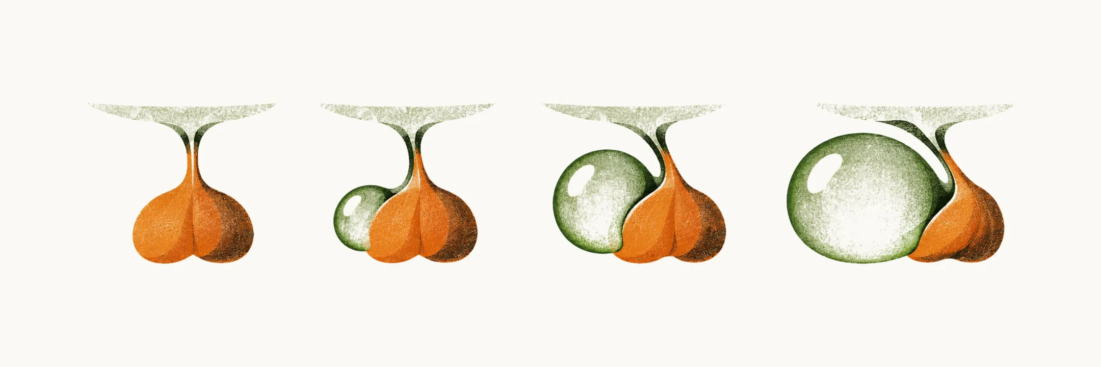
The pituitary sits in the sella, the bony cup that holds the gland, and the stalk runs up from it to the hypothalamus. Several masses besides the pituitary adenoma arise in this region. They come from different tissues: remnants of Rathke’s pouch, the meninges, cancers elsewhere in the body, and the hypothalamus and optic pathways.
A mass takes its identity from the tissue it started in, not from the gland’s hormone cells. Several of these tissues lie close to the optic chiasm, the stalk and the hypothalamus, so a mass from them can press on vision and on the hormone supply. The useful question to ask of any sellar mass is therefore which tissue it started in.
Where does Rathke’s pouch come into it?
The first tissue in the list is the one that builds the front of the gland, so its remnants come next. The gland has two embryological parts, and the front part is built from Rathke’s pouch, while the back part comes from the brain. Two of the masses below arise from the pouch: craniopharyngiomas and Rathke’s cyst. A craniopharyngioma is thought to come from epithelial remnants of the pouch or of the craniopharyngeal duct, and a cyst from a remnant that fails to close.
Why is a benign mass dangerous here?
Craniopharyngiomas are benign, cystic masses, so the question is why a benign tumour matters at all. “Benign” describes how the cells behave. The harm comes from the position, next to the optic pathways and the hypothalamic-pituitary axis.
The position follows from the embryology. The pouch extends down to fuse with the infundibulum and forms the craniopharyngeal duct. That is why craniopharyngiomas are derived from Rathke’s pouches near the pituitary stalk. The Rathke’s pouch-derived tumours mostly extend to the suprasellar cistern.
Who gets a craniopharyngioma, and what shows first?
A mass that sits at the stalk raises the question of who gets it and when. Craniopharyngiomas are diagnosed in both children and adults. The age distribution is bimodal, with one peak in children aged 5-14 years and another in adults aged 50-75 years.
In younger patients, the clinical complaints often come from increased intracranial pressure: headache, vomiting, papilledema and hydrocephalus.
How does the mass take away vision and hormones?
The next question is what the mass does to vision and to the hormones. Craniopharyngiomas present with a visual field deficit, headache, and a variable degree of hypopituitarism. The visual loss comes from the mass compressing and stretching the optic nerves near the chiasm.
The gland fails for a different reason. Hypothalamic signals reach the gland down the stalk in a portal system. A mass that damages the hypothalamus and stalk cuts the gland off from the signals it needs. This explains why hypopituitarism is documented in up to 90% of craniopharyngioma cases at diagnosis.
Why does diabetes insipidus appear?
The same damage to the hypothalamus and stalk reaches the water-balance hormone. ADH is made in the hypothalamus and stored in the posterior pituitary. A mass that damages this supply reduces ADH, and the kidney cannot keep water. At diagnosis, diabetes insipidus is documented in about 10% of craniopharyngioma cases.
What does the hypothalamus add?
The hypothalamus is the part the mass presses on, and it helps control appetite and sleep. This explains the other craniopharyngioma symptoms: weight gain accompanied by metabolic syndrome, sleep disturbances and cognitive changes.
How is a craniopharyngioma treated?
Craniopharyngioma treatment is usually based on transcranial or transsphenoidal surgery with post-operative radiotherapy. The next question is about the cyst that comes from the same pouch.
What is a Rathke’s cyst?
The cyst comes from the same pouch, and it also sits at the stalk. As the gland grows, the pouch lumen normally closes and leaves a thin residual cleft. If the developmental obliteration of the pouch fails, a Rathke’s cyst can form. It is a small cyst that can compress the stalk.
What does a Rathke’s cyst cause?
A cyst that presses on the stalk causes the symptoms that come next. Its main clinical symptoms are diabetes insipidus and hyperprolactinemia. The diabetes insipidus follows because compression of the stalk and posterior lobe reduces the ADH supply, as above. The hyperprolactinemia follows because interrupting dopamine flow down the stalk raises prolactin.
Why are some sellar meningiomas hard to tell from adenomas?
Pressure from a cyst and from a sellar mass can look alike, so the next question is which other masses look alike. It is difficult to distinguish a non-functioning pituitary adenoma from a meningioma arising in the sella turcica. Both sit in the sella and press on the same gland and optic structures, so their pressure effects overlap. The difference is the tissue of origin, and imaging has to establish it, because each tissue has its own MRI pattern.
A meningioma arises from the dura over the sella. Meningiomas may cause compressive symptoms in the patient, and compression of the optic nerve is one way they threaten vision.
How do cancers reach the pituitary?
The meningioma comes from the meninges, but a different source, a cancer elsewhere in the body, can also reach the gland. Pituitary metastases occur in 3% of cancer patients. They mostly occur in the posterior pituitary gland. The posterior lobe and the infundibulum have a direct arterial blood supply, which makes them accessible to circulating tumour cells.
A metastasis there cuts the ADH supply, so it can cause diabetes insipidus. About half of pituitary masses come from breast cancer.
What happens when the hypothalamus itself grows a mass?
A metastasis to the posterior lobe comes from elsewhere, but these masses come from the hypothalamus itself. Hypothalamic hamartomas and gangliocytomas may arise from oligodendrocytes, astrocytes or neurons. Hypothalamic hamartomas are built from ectopic neuronal and glial tissue, which is why the cells can be of either kind.
These tumours can over-produce some hypothalamic hormones, namely GnRH, GHRH or CRH. GnRH raises FSH and LH, so a mass that secretes it starts the gonadal axis early. Children with the GnRH-producing type manifest with precocious puberty, psychomotor delay and laughing-associated seizures. The seizures are thought to arise from the lesion’s own epileptic activity, and the mechanism is not well understood.
Why do visual-pathway gliomas depend on age?
Hypothalamic tumours can also start in the visual pathway. Hypothalamic gliomas and optic gliomas mostly occur in children. In children they are typically low-grade pilocytic astrocytomas that often behave benignly. They are more aggressive in adults, where the sporadic tumours are aggressive, high-grade malignancies.
Which cells do the gland’s own tumours come from?
Every mass so far starts in a tissue other than the gland’s hormone cells. Adenomas arise from the gland’s own cells, and none of these other masses does. That is what sets the pituitary adenoma apart. So which cell types of the gland give rise to adenomas, and how common are they?