
The pituitary gland sits in the sella, with the optic chiasm just above and the pituitary stalk running down to it from the hypothalamus. Non-functioning sellar tumours make very little hormone or none. An adenoma that makes no hormone gives no hormonal warning, so these tumours cause no clinical change in the patient and no hormone syndrome draws attention to them. They tend to be recognised through the pressure they exert.
Pressure reaches the nearest structures first: the chiasm, the stalk and the normal gland. Pressure on the stalk cuts the dopamine that holds prolactin back. The raised prolactin then blocks the pulsed release of GnRH that drives FSH and LH, so the gonads go quiet. These links explain the three usual signs: visual disturbance, hyperprolactinemia and hypogonadism.
How large does it have to be before anyone notices?
A silent tumour gives no hormonal warning, so the first sign is pressure, and the question is how much pressure is needed. NFPAs are mostly macroadenomas at diagnosis. They cause clinical effects only when they are big enough to produce a compression effect. NFPAs are usually asymptomatic during the first phases. A larger mass presses harder on what is next to it. They become symptomatic when they are big enough to press on the optic chiasm.
Who gets these tumours, and how common are they?
Being found only once large raises the question of who gets them. NFPAs are the most common sellar mass after prolactin-producing tumours of the sellar region. They affect males and females equally, and they are mostly diagnosed at age 40-50.
The gonadotropinoma is more prevalent in males. It is believed, however, that gonadotropinoma is extremely under-estimated in females.
What cell does a silent tumour come from?
Because these tumours are common and mostly silent, the next question is which cell they come from. For an adenoma, the cell type it came from sets the hormone picture. Based on histochemical studies, most NFPAs originate from gonadotropin cells or null cells. A tumour from a gonadotroph keeps some of the cell’s hormone-making machinery. Null cells produce no hormone.
What hormone does it leak, if any?
If the cell is a gonadotroph, the next question is what hormone, if any, it releases. Non-functioning tumours usually make small amounts of intact gonadotropins such as FSH. FSH and LH are each a common alpha subunit joined to a hormone-specific beta subunit, and the join is what makes them active, so unjoined parts are not working hormone.
These tumours also usually make uncombined subunits: LH-β, FSH-β and the glycoprotein α subunit. Free beta subunits are largely degraded inside a normal cell when they are not joined, presumably because of improper folding. A tumour that releases them free is therefore releasing parts that normal assembly would have kept.
When a non-functioning gonadotropin-derived neoplasm is stimulated with TRH, it shows an atypical increase of tumour-derived gonadotropin hormones and subunits.
Can leaked gonadotropins reach the ovary?
Leaked FSH and LH are what the ovary answers to. Very rarely, NFPAs cause menstrual disturbances and ovarian hyperstimulation. In a reported case the estradiol was high while FSH was not suppressed and LH was suppressed, which is consistent with an FSH-producing tumour acting on the ovary. In these cases the patients mimic polycystic ovary syndrome (PCOS), with a high level of estrogen production. The resemblance lies in a shared picture of cysts and high estrogen, not in a shared cause.
What does the pressure do to the rest of the gland?
The pressure, not the hormone, tends to give them away, so the next question is what it does to the normal gland. The most common manifestations of NFPAs are:
- compression effects, which are visual disturbances and headache;
- hypopituitarism, with loss of ACTH, TSH and gonadotropins;
- hyperprolactinemia.
The pattern of visual field loss depends on the site of compression, so the visual signs vary. A mass can squeeze the normal gland, leaving it under-active, which explains the hypopituitarism. Dopamine from the hypothalamus holds prolactin back, and cutting its flow down the stalk raises prolactin. Together this is why NFPAs manifest with visual disturbances, hyperprolactinemia and hypogonadism.
How does a squeezed stalk lower the sex hormones?
The stalk carries dopamine down to the gland, so the next question is how squeezing it lowers the sex hormones. Hypogonadism comes from compression of the pituitary stalk and the resulting hyperprolactinemia. Compression lowers the dopamine that reaches the gland, so prolactin rises.
The resulting hyperprolactinemia blocks the pulsatile release of GnRH. It does so by suppressing the hypothalamic neurons that drive that pulsed release, so the gonadotropin signal falls.
How do you tell a prolactin-making tumour from a squeezed stalk?
Raised prolactin can have two sources, so the next question is how to tell them apart. In a radiological macroprolactinoma, the prolactin level normally rises with tumour size, because the tumour itself secretes it. If the prolactin level does not fit the tumour size, consider a second explanation: the tumour is not functioning, and the hyperprolactinemia comes only from pituitary stalk compression. A typical example is a macroprolactinoma with a prolactin of 100-200 ng/ml that does not correlate with tumour size.
The two sources work differently. A squeezed stalk gives raised prolactin through decreased dopamine, while a prolactin-making tumour gives it through increased secretion. The squeeze removes a brake, so the rise is smaller. A true macroprolactinoma usually raises prolactin above 200 ng/ml, whereas hyperprolactinemia from stalk compression is usually below about 100 ng/ml. A value in between is indeterminate.
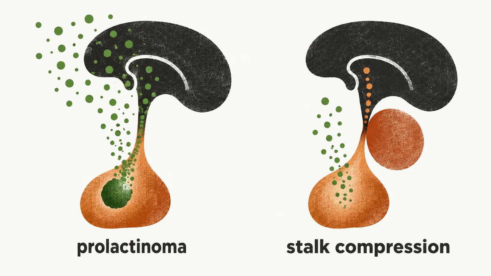
Why does the difference change the treatment?
Once the two are told apart, the next question is why that changes what is done. The two scenarios must always be differentiated because of the difference in treatment. Drugs for a prolactin-making tumour act on the prolactin brake, so in a true macroprolactinoma pharmacological treatment is used. Dopamine agonists will not shrink a non-functioning tumour that is causing pituitary stalk compression, although prolactin levels will decrease. A drug would therefore lower the prolactin without removing the mass that is squeezing the stalk. In stalk compression, the patient is operated on.
Why is it easy to miss after menopause?
The gonadotropin signal of the tumour looks much like the one at menopause, so the next question is what menopause does to it. GnRH raises FSH and LH. Estradiol restrains the pituitary’s response to GnRH, more for FSH than for LH. During and after menopause the lack of estrogen production removes that restraint, so FSH and LH are hypersecreted, which is the same characteristic as gonadotropinoma. Because the menopausal rise looks like the tumour’s rise, the tumour’s gonadotropin is easily put down to menopause. Under-diagnosis, especially in women of menopausal age, is very common.
What does it look like at each age and sex?
The same signal looks different across life, and the clinical manifestation of gonadotropinoma differs based on age and sex.
- Males before puberty: precocious puberty.
- Males after puberty: erectile dysfunction and hypoactive desire.
- Females before puberty: precocious puberty.
- Females before menopause: amenorrhea and menstrual irregularities.
- Females after menopause: the hormonal effect is masked by the signs and symptoms of menopause itself.
At puberty the gonads become more responsive to FSH and LH, so a tumour that releases them before puberty can supply that signal early. After menopause FSH and LH are already raised, so the tumour’s extra hormone adds to a raised baseline and the signs blur. In both sexes, visual disturbance is a common way a gonadotropinoma comes to attention, because a mass growing upward presses on the optic chiasm.
If these tumours are found by pressure, where do they fit among all the masses that can occupy the sella, and where should a student start?