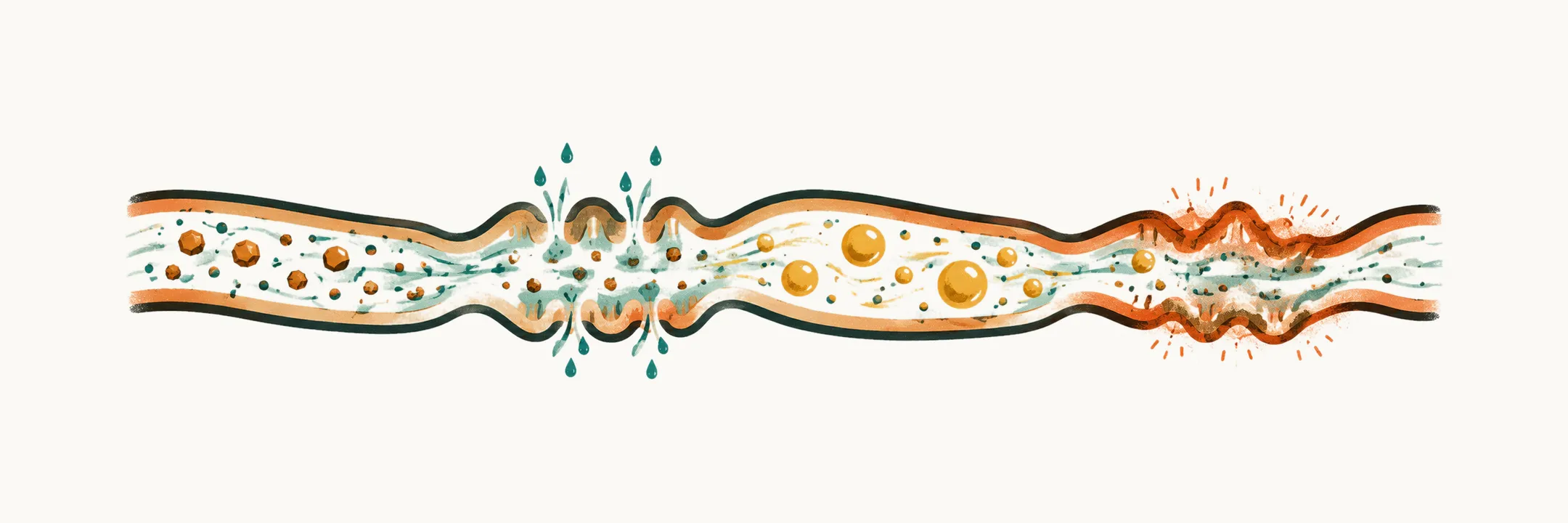
Chronic diarrhea in children is defined by duration first and explained by mechanism second. Duration comes first because it is what you learn at the first visit, and it separates chronic diarrhea from the acute illness that usually precedes it. Mechanism comes second because it decides which part of the work-up to follow, and so which diagnosis is even possible.
When does diarrhea count as chronic?
The thresholds in pediatric practice sit on a continuum that begins with acute gastroenteritis.
- Acute diarrhea lasts less than 7 days, and in practice not longer than 14 days. In Europe it is viral in the great majority of children, and it is managed by replacing fluid.
- Prolonged diarrhea lasts 7 to 14 days. Often it is an acute illness resolving slowly. But this is already the point at which a non-infectious cause may be declaring itself.
- Chronic diarrhea lasts 14 days or more.
Some pediatric texts reserve the word chronic for 4 weeks or more, and sources use the two thresholds, 14 days and 4 weeks, interchangeably. The disagreement is real, but the clinical consequence is the same: by then the differential has changed completely. So the exact threshold matters less than the shift it marks.
What is that shift? Acute diarrhea in children is overwhelmingly infectious. Chronic diarrhea is functional in many children, and it is also where malabsorption, immune-mediated mucosal disease and congenital defects live. Rehydrating the child settles none of these, because the problem is the mucosa or the digestion, not lost fluid.
One older label belongs here. Intractable diarrhea is a historical term for severe diarrhea of infancy that does not respond to enteral feeding and requires parenteral nutrition. It describes a group of congenital enteropathies rather than a duration.
How does the gut end up with loose stool?
Water movement across the intestinal mucosa normally follows solute: water goes where dissolved particles go. Sodium-coupled uptake of sugars and amino acids pulls water out of the lumen and into the body. The colon then absorbs nearly all of what remains, so the stool that is passed is mostly formed.
The stool turns loose when that balance fails. Anything that adds an unabsorbed solute to the lumen, drives secretion, damages the mucosa or blocks fat digestion produces loose stools. Each mechanism does it differently, and the difference is visible at the bedside and in the stool. The four mechanisms are osmotic, secretory, fat malabsorption and inflammatory.
How do the osmotic and secretory mechanisms differ?
In the osmotic mechanism an unabsorbed solute holds water in the lumen. Pediatric examples are lactose or other disaccharide intolerance, excessive fruit juice or fructose, and some malabsorptive states. Because the solute is the cause, the stools stop when the child fasts: with no food, no solute is left to hold the water.
When the solute is a carbohydrate, the stool is acidic, with a pH below about 5.5, and the reducing substances are positive, because unabsorbed sugar is present in the stool. A greater than 100 mOsm/kg also fits an osmotic mechanism.
In the secretory mechanism the mucosa actively secretes chloride and water. This draws fluid from the blood into the lumen whatever the child eats, so the stools continue during fasting and the volumes are large. The stool osmotic gap is below 50 mOsm/kg. Examples are enterotoxin-driven diarrhea, , and laxative or osmotic-drug ingestion. As an isolated mechanism it is rare in children.
Why does a gap separate the two? Stool is close to plasma in osmolality, and the gap is the part of that osmolality that sodium and potassium do not account for:
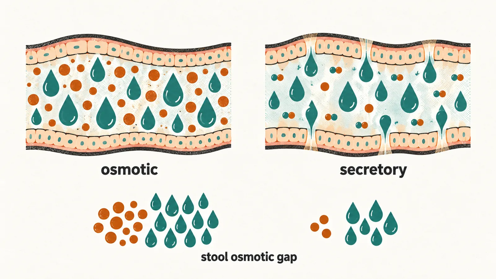
with sodium and potassium in mmol/L. The electrolytes are doubled to allow for the anions that go with them. A high gap means something else is in the stool, an unabsorbed solute, and that points to an osmotic cause. A low gap means the osmolality is made up of electrolytes, which points to active secretion. A gap between 50 and 100 mOsm/kg is intermediate and does not settle the question on its own.
What do fat malabsorption and inflammation add?
Not every loose stool is a watery one. Two mechanisms leave their own marks.
Fat malabsorption (steatorrhea). Fat needs pancreatic enzymes and bile salts to be digested, and an intact mucosa to be absorbed. When any of these fails, fat is not digested or not absorbed, and the unabsorbed load carries water with it. The stools are pale, bulky, greasy and foul-smelling, sometimes with visible oil. The osmotic gap is high. Causes that belong together for this reason are:
- pancreatic exocrine insufficiency, with cystic fibrosis the most important cause;
- chronic pancreatitis;
- bile-salt deficiency;
- mucosal disease such as celiac disease.
Because the problem is pancreatic or mucosal, the investigation differs from the rest. With pale, greasy, offensive stools and a child who is not growing, the next tests are for pancreatic exocrine insufficiency and a sweat chloride test for cystic fibrosis. A sweat chloride of 60 mmol/L or more is diagnostic of cystic fibrosis. A value below 30 mmol/L makes cystic fibrosis unlikely.
Inflammatory mechanism. Inflamed mucosa leaks blood, mucus, protein and water. That explains the blood and mucus in the stool. Inflammation of the rectum and distal bowel is thought to produce urgency and tenesmus. Inflammatory mediators reach the blood, so CRP or ESR are raised. Neutrophils enter the inflamed bowel and release calprotectin, so fecal calprotectin, a stool protein released by neutrophils, is raised. Examples are inflammatory bowel disease, cow’s milk protein allergy, eosinophilic gastroenteritis and infections.
Are the mechanisms ever pure?
Pure mechanisms are the exception. Most children with chronic diarrhea have a mixed picture. One disease can damage the mucosa and also leave solute behind. Examples are an osmotic load from malabsorbed carbohydrate layered on mucosal inflammation, and fat malabsorption with an osmotic component.
This limits the fasting test. The fasting response is a bedside observation, not a laboratory test. Withholding food from an already faltering child to prove a mechanism costs more than it gives.
Which mechanism is likely at this age?
Mechanism explains how the stools are produced. Age says which diseases are likely to produce them. Age narrows the list faster than any single symptom, so a chronic diarrhea work-up always starts with how old the child is.
- First month: congenital and monogenic enteropathies, which are inherited defects of the intestinal lining, such as and . Also congenital disaccharidase defects and , congenital chloride diarrhea, primary immunodeficiency, intestinal lymphangiectasia (dilated lymph vessels in the bowel wall) and very early onset IBD.
- 1–6 months: cow’s milk protein allergy is the leading cause. Congenital enteropathies become less common than in the neonatal period.
- 1–5 years: functional (toddler) diarrhea is the single most common cause. Celiac disease appears after gluten is introduced. Cystic fibrosis–related pancreatic insufficiency and cow’s milk protein allergy persist.
- Older child and adolescent: IBD becomes a major consideration, and irritable bowel syndrome is common. Celiac disease often presents without diarrhea at all, with short stature, anemia or dermatitis herpetiformis (itchy blistering skin rash).
These bands are tendencies rather than rules. A congenital enteropathy can present late, and IBD can present in the first year of life. So the age table narrows the questions to ask rather than answering them. Benign patterns dominate the young end of it.
If age and stool pattern now point toward a mechanism, in what order should a child with chronic diarrhea be questioned, examined and tested?