
Acute promyelocytic leukemia (APL) is a subtype of acute myeloid leukemia (AML). It is uncommon, about 7% of childhood AML. Yet it looks least like any other childhood leukemia in the way it is treated, and it can kill a child by bleeding before any chemotherapy has been given. Why is it so different?
What makes APL different from other childhood leukemias?
Like other AML, APL is a clonal proliferation of myeloid precursor cells. They accumulate in the marrow and displace normal blood-cell production.
What sets APL apart is a translocation. The t(15;17) fuses PML on chromosome 15 with RARA on chromosome 17, and the product is the PML::RARA fusion protein. One stuck cell, held by this one protein, explains both the danger and the cure. Why is the cell stuck?
Why does the cell stop at the promyelocyte stage?
A neutrophil is made by step-by-step ripening. A myeloid precursor passes through the promyelocyte stage and then goes on. RARα is a retinoic acid receptor, and when it binds retinoic acid it switches on genes that carry the cell onward.
The PML::RARA fusion acts as a transcriptional repressor instead. It is thought to sit on those same genes and recruit repressing partners, so the genes the cell needs to move on stay switched off. The precursor is held at the promyelocyte stage. Cells cannot mature, so they accumulate.
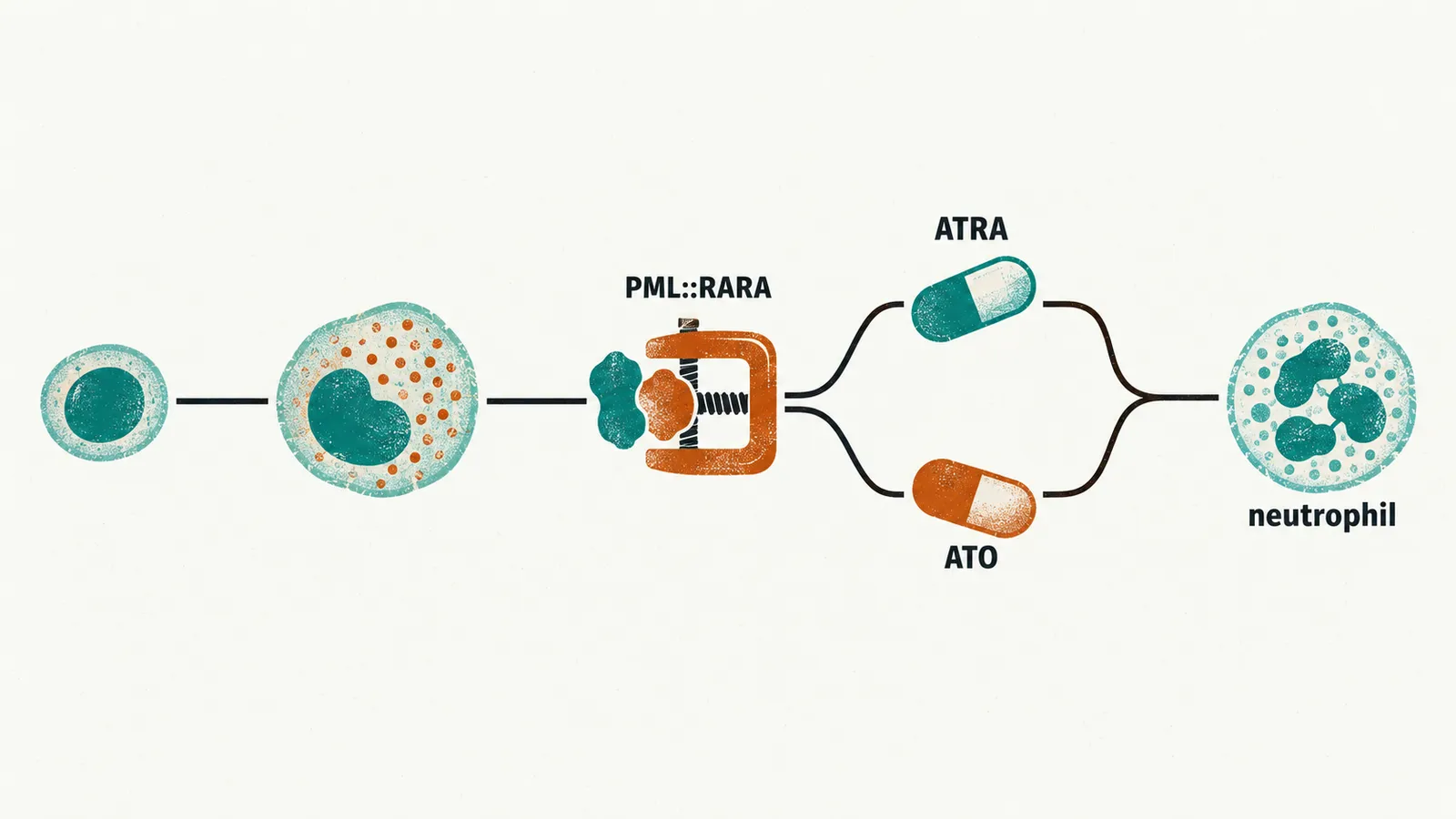
They accumulate full of granules and Auer rods, because the block arrests them at a granule-rich stage. The granules and Auer rods give APL its morphological signature, so suspicion can arise from the slide before the translocation result is back. But these granules are not harmless.
Why does APL cause bleeding and clotting together?
Almost every child with APL has a coagulopathy at diagnosis. Part of this comes from the stuck cells themselves.
The promyelocyte granules and Auer rods release procoagulant material. Tissue factor on the blast surface activates the coagulation cascade; the disturbed signaling of the fusion is thought to raise it. At the same time, annexin II drives fibrinolysis, the breakdown of clots.
The result is a picture of disseminated intravascular coagulation: clotting activated throughout the circulation, with platelets and clotting factors used up. The findings follow from this:
- thrombocytopenia, as platelets are used up;
- prolonged prothrombin and partial thromboplastin times, as clotting factors are depleted;
- raised D-dimers, as clots are broken down;
- low fibrinogen, as it is consumed.
The coagulopathy can worsen in the first days of treatment. Because clotting and fibrinolysis are on together, bleeding and thrombosis appear together. Early deaths in APL are usually hemorrhagic, because the loss of platelets and clotting factors makes bleeding the dangerous side. So what should be done when the diagnosis is suspected?
What is done on suspicion, before the diagnosis is confirmed?
The cost of waiting is bleeding. So any child with new acute leukemia and abnormal clotting is treated as if APL until cytogenetics or molecular testing say otherwise.
- Platelets and fibrinogen are replaced before bleeding starts, because both are consumed and both are needed to form clots.
- ATRA is started on morphological suspicion, rather than after the translocation result. Waiting would delay a drug that matures the very cells making the procoagulants.
- Lumbar puncture at diagnosis waits until the coagulopathy has resolved, because an invasive procedure is dangerous while clotting is failing.
Some features mark a greater risk of coagulopathic complication: a high white cell count, a low platelet count, abnormal coagulation studies, a high body mass index, and FLT3 internal tandem duplication, a duplicated stretch within the FLT3 gene. Once clotting is covered, what actually treats the disease?
How can APL be treated without chemotherapy?
APL can be treated without conventional cytotoxic chemotherapy, which is true of almost no other acute leukemia. The reason is that the driver can be removed directly.
The modern backbone is ATRA combined with arsenic trioxide (ATO). Chemotherapy is reserved for high-risk presentations such as a high white cell count. The two drugs attack the same fusion protein in different ways.
- ATRA binds the RARα portion and releases the transcriptional repression imposed by the fusion. The promyelocytes resume the maturation program they were arrested in and differentiate into neutrophils rather than being killed.
- ATO binds the PML portion. It triggers degradation of the PML::RARA protein through sumoylation (tagging with small ubiquitin-like modifier proteins) and the proteasome.
Differentiation and degradation together remove the driver. That is why most patients are cured without classical cytotoxic chemotherapy. But a drug that matures the cell does so with consequences.
What does the treatment itself do to the child?
Treatment brings two toxicities: one from the maturation produced, and one from arsenic trioxide itself.
Differentiation syndrome comes from the same maturation that makes therapy effective. Cells are thought to release cytokines as they differentiate, and capillaries leak. Fluid leaving the capillaries is behind several of the features: fever, dyspnea, weight gain, pleural or pericardial effusions, pulmonary infiltrates and hypotension. It is treated with dexamethasone, started at first suspicion.
Arsenic trioxide adds QT prolongation, a lengthening of the QT interval on ECG. Electrocardiograms and potassium and magnesium monitoring are part of the treatment plan. How well does all this work, and for which children?
Do children and adults differ, and how good are the results?
A randomized comparison of ATRA plus ATO against ATRA plus chemotherapy, published in 2013, established the chemotherapy-free combination in adults with low- and intermediate-risk disease.
is defined by a white cell count of 10 × 10⁹/L or above. Children present at or above that threshold more often than adults do. They also present more often with the , which is often associated with higher white cell counts.
The Children’s Oncology Group AAML1331 trial gave tretinoin and arsenic trioxide without cytotoxic chemotherapy to standard-risk children. High-risk children received the same two drugs plus 4 doses of idarubicin during induction.
| Risk group | Treatment in AAML1331 |
|---|---|
| Standard risk | Tretinoin and arsenic trioxide |
| High risk | Tretinoin and arsenic trioxide plus 4 doses of idarubicin in induction |
The trial reported 3 relapses and 1 death among 154 children, with no significant difference in outcome between risk groups. So with tretinoin- and arsenic trioxide-based therapy, high-risk disease no longer carries a clearly worse outcome.
Cure rates for APL with ATRA and ATO are high, above 90% in trial data for adults. The same approach has transformed pediatric APL from the most lethal subtype, where bleeding was often fatal, to one of the most treatable. Relapse after remission induction occurred in about 2–4% of children in pediatric trials. What makes this possible is a driver that two drugs can remove.
What happens when there is no removable driver?
Neuroblastoma has no single driver of that kind to take out. Its treatment is built on age, stage and biology. It is a different problem from the leukemias in almost every respect. If neuroblastoma has no single driver to remove, how do its clinical signs point to the diagnosis, and how does age shape its treatment? Neuroblastoma answers that.