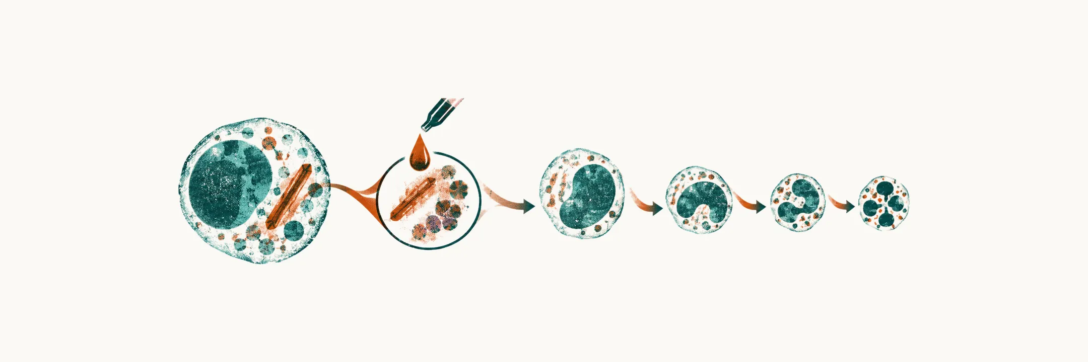
Acute myeloid leukemia (AML) is a malignant clonal proliferation of myeloid precursor cells. These are the marrow cells that normally mature into granulocytes, monocytes, red cells and platelets. When one clone stops maturing and piles up, the marrow can no longer make these cells in the normal way.
AML is the second most common childhood leukemia, and a distant second. It accounts for 15–20% of pediatric acute leukemias, and the remaining 80% or so are acute lymphoblastic leukemia (ALL). One AML subtype, acute promyelocytic leukemia (APL), behaves differently from every other acute leukemia. Its promyelocytes are held short of maturation by a single fusion protein, and its treatment makes them mature rather than killing them.
Does the child look different from a child with ALL?
As in ALL, the presenting signs come from marrow whose normal production is displaced by blasts. Anemia, neutropenia, thrombocytopenia and hepatosplenomegaly can be the opening picture in either disease.
Some features point toward the myeloid side. Monocytic differentiation (FAB M4 and M5) is associated with gum hypertrophy, with skin infiltration and with a higher rate of CNS involvement. AML can invade tissue outside the marrow, such as the meninges, brain, testes, ovaries or skin, and this is one way these findings could arise.
A myeloid sarcoma (chloroma) is an extramedullary mass of myeloid blasts, a tumor outside the marrow. It can appear before the marrow is visibly involved. The clone is already present before it fills the marrow, so a mass may be the first place it shows.
Why can a high white cell count be an emergency by itself?
White cell counts run higher in AML than in ALL. Higher counts bring a separate emergency: leukostasis, sludging of blasts in small vessels of the brain, lungs and other organs.
Why does sludging happen? When very many blasts circulate, they can obstruct small vessels. Proposed contributors are that myeloid blasts are less pliable than mature granulocytes or lymphoblasts, that they stick to the vessel wall, and that they damage the endothelium. This is thought to explain why the signs are mostly neurologic and respiratory: headache, confusion, dyspnea, visual disturbance or priapism.
Because blasts in the vessels are the problem, leukostasis needs urgent cytoreduction (lowering the blast count with leukemia-directed treatment) rather than waiting for a definitive diagnosis.
How is the lineage decided?
Clinical clues suggest AML, but none settles it. Lineage is decided in the laboratory, not at the bedside. This matters because the treatment backbones of AML and ALL differ.
Three laboratory tools are used together, and each reads a different property of the blast:
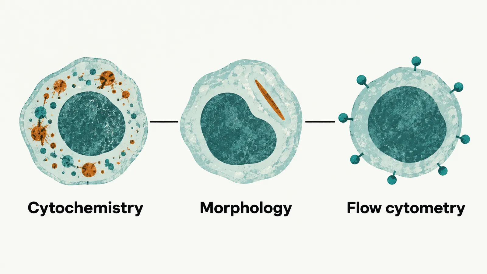
- Cytochemistry stains blasts for enzymes. Myeloperoxidase (MPO) positivity indicates AML. It is the single most useful stain, because MPO is present in myeloid blasts and essentially absent in lymphoid ones.
- Morphology is the appearance of blasts under the microscope. Auer rods indicate AML. They are needle-shaped cytoplasmic inclusions of fused azurophilic granules, and they are pathognomonic of myeloid lineage, seen in no other lineage. They are often abundant in APL.
- Flow cytometry reads the surface markers carried by individual cells. The myeloid markers are CD13, CD33 and CD117, present in place of the B- or T-lineage markers of ALL.
Which kind of AML is it?
Once the lineage is myeloid, the next question is which kind of AML. Two classifications answer it.
The older French-American-British (FAB) scheme divided AML by morphology into M0 to M7:
- M0: minimally differentiated
- M1: myeloblastic without maturation
- M2: myeloblastic with maturation
- M3: promyelocytic, the subtype APL falls into
- M4: myelomonocytic
- M5: monocytic
- M6: erythroleukemia
- M7: megakaryoblastic
FAB remains useful vocabulary, and M4 and M5 are the monocytic types met above.
The current WHO classification is built instead on defining genetic abnormalities, because chromosomal and molecular abnormalities are important diagnostic and prognostic markers. They are:
- the fusion genes PML::RARA, RUNX1::RUNX1T1 and CBFB::MYH11
- KMT2A rearrangement
- NPM1 mutation
- a myelodysplasia-related category
FLT3 mutation is not defining. It still carries prognostic weight and now directs therapy: certain FLT3 variants are associated with a high risk of relapse, and finding them may allow targeted therapy. form a separate WHO category.
How is AML treated?
Because the genetics carry the prognosis, they also shape the treatment. Pediatric AML treatment is more intensive and shorter than ALL treatment, with less emphasis on prolonged maintenance. The chemotherapy runs in sequence: induction, then consolidation.
For induction, the long-standing backbone is daunorubicin with cytarabine. This is the “7 + 3” schedule: 7 days of cytarabine and 3 days of anthracycline. Pediatric regimens adapt it.
Consolidation is high-dose cytarabine blocks, usually two or more. These blocks do most of the work of preventing relapse. CNS prophylaxis with intrathecal therapy is also given, because AML can invade the meninges and brain, although it does so less often than ALL.
Other elements depend on the child’s risk and disease biology, so the biology picks the add-on:
- allogeneic hematopoietic stem cell transplantation, from a donor, for high-risk disease in first remission and for many children with relapsed disease
- gemtuzumab ozogamicin, an anti-CD33 antibody-drug conjugate (an antibody that carries a cytotoxic drug to cells bearing its target), used in CD33-positive disease
- FLT3 inhibitors, used where the disease is FLT3-mutated
Reported 5-year overall survival for childhood AML is now in the region of 65–70%. It varies widely by subtype, which fits the weight the defining genetics carry. Relapse remains the main cause of treatment failure.
Why does Down syndrome change the treatment?
Children with Down syndrome have a substantially increased risk of acute leukemia, specifically of acute megakaryoblastic leukemia (AML M7). This category is now called myeloid leukemia of Down syndrome. It is defined by blasts with GATA1 variants in a child with trisomy 21.
GATA1 is needed for normal development of megakaryocytes. The leukemia variants leave a truncated factor with decreased activity, and the combination of trisomy 21 and a damaged GATA1 is the setting in which this clone is seen.
Outcome with chemotherapy is generally excellent. A proposed explanation is that GATA1 variants increase sensitivity to cytarabine by lowering cytidine deaminase. Yet these children experience significantly greater treatment toxicity. Reduced-intensity regimens are therefore used to obtain the same result with less harm.
The lesson is not confined to Down syndrome: the biology of a myeloid leukemia decides how much treatment a child can safely be given. In APL, biology decides which drugs are used at all. In that subtype a single fusion protein holds the promyelocytes back, so what happens when treatment makes them mature? That is the question of Acute Promyelocytic Leukemia in Children.