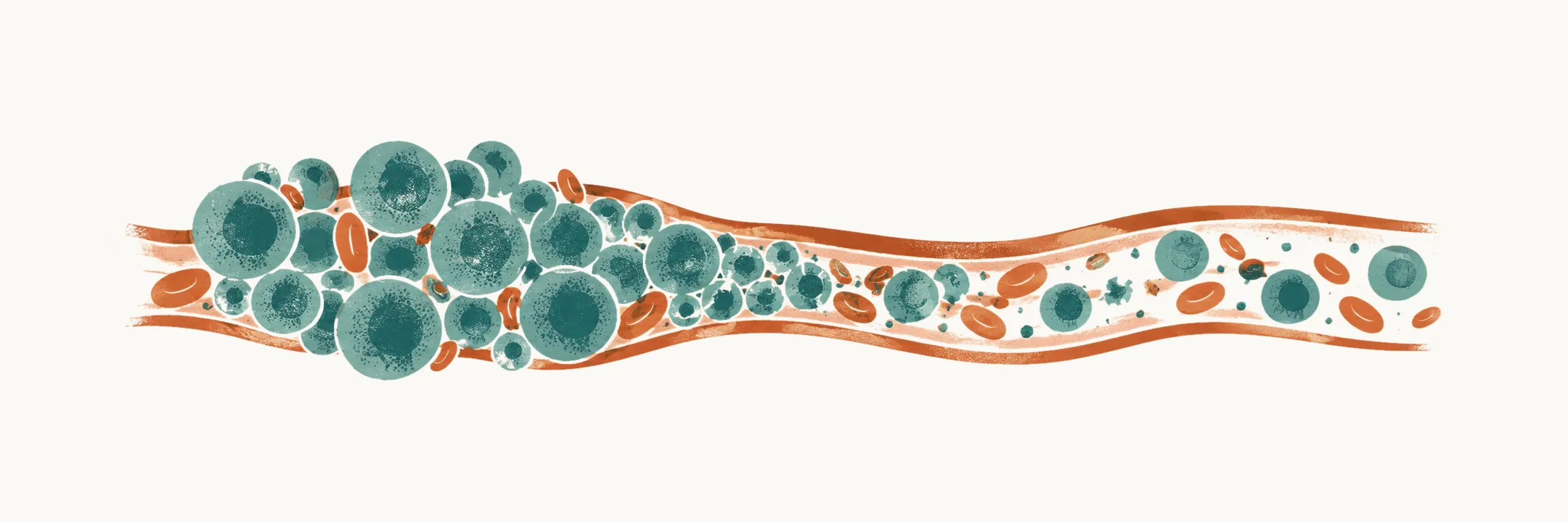
Several complications of childhood leukemia and neuroblastoma, and of their treatment, can become life-threatening within hours. A child can die of one of them before the cancer itself has been treated.
Why is that so? Three simple pictures explain it. A leukemic clone is a very large, fast-turning population of cells. If it dies all at once, its contents flood the blood. If it grows too large, it crowds the circulation and disturbs clotting. Second, a child is a small space, so a mass in the chest or beside the spine presses on something that has no room to give. Third, chemotherapy removes neutrophils, so infection can run ahead of any warning.
That is why these conditions are recognized from history and examination, rather than from a test that arrives in time. Each turns on one or two decisions: fluid, antibiotics, position, cytoreduction, steroids. Six of them account for most of the early danger, and they fall into three groups.
Which six, and how do they group?
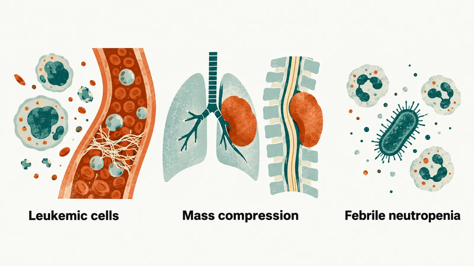
The first group is problems caused by leukemic cells themselves: tumor lysis syndrome, leukostasis and the coagulopathy of acute promyelocytic leukemia (APL). These cover what cells release when they die in large numbers, how they crowd small vessels, and what they do to clotting.
The second group is a mass pressing on a vital structure: a mediastinal mass compressing the airway, and spinal cord compression.
The third group is infection when treatment has left too few neutrophils: febrile neutropenia.
The first group begins with what happens when the clone dies.
What happens when a large clone dies at once?
Tumor lysis syndrome (TLS) is the metabolic consequence of leukemia cells dying in large numbers at once. It usually follows the start of chemotherapy, when a large, rapidly dividing population is destroyed together. It can also begin spontaneously in bulky disease.
The breakdown of such a population releases potassium, phosphate and nucleic acids into the blood. The kidneys cannot clear them as fast as they arrive. Four abnormalities follow, and the calcium falls secondarily because phosphate binds it in the blood:
- hyperkalemia
- hyperphosphatemia
- hyperuricemia
- secondary hypocalcemia
Together they cause acute kidney injury, arrhythmia, seizures and death. Because the risk can be estimated from tumor burden before any blood result is abnormal, TLS management is anticipatory.
How is tumor lysis recognized, and when does it count as clinical?
Laboratory TLS is defined by at least two of the four abnormalities in the same 24-hour period, from 3 days before to 7 days after treatment starts. One abnormal value on its own may not come from TLS. The thresholds in children are:
| Abnormality | Threshold in children |
|---|---|
| Uric acid | ≥8.0 mg/dL (476 µmol/L), or ≥25% increase from baseline |
| Potassium | ≥6.0 mmol/L, or ≥25% increase from baseline |
| Phosphate | ≥6.5 mg/dL (2.1 mmol/L), or ≥25% increase from baseline |
| Calcium | ≤7.0 mg/dL (≤1.75 mmol/L), or ≥25% decrease from baseline |
The phosphate threshold is the one that differs from adult practice, where it is 4.5 mg/dL (1.45 mmol/L).
Clinical TLS is laboratory TLS plus at least one of these: creatinine ≥1.5 times the age-adjusted upper limit of normal; arrhythmia or sudden death; seizure. So the clinical form adds an organ consequence to the laboratory picture.
The definition fixes the numbers; the management fixes the order of events.
How is tumor lysis prevented and treated?
The management has four parts.
- Intravenous hydration without potassium. Fluid keeps flow through the kidney up, and potassium is left out because the clone is already releasing it.
- Frequent measurement of potassium, phosphate, calcium, uric acid, creatinine and lactate dehydrogenase, because the values can change quickly.
- Uric acid-lowering drugs, allopurinol or rasburicase, according to risk. Rasburicase is not given to a child with G6PD deficiency, in whom it causes hemolysis and methemoglobinemia.
- Dialysis when hyperkalemia, hyperphosphatemia or renal failure are refractory.
Asymptomatic hypocalcemia is not corrected reflexively. Added calcium can precipitate with phosphate and worsen renal injury, so the aim is to relieve symptoms rather than to treat the number.
Tumor lysis is a problem of too many cells dying. What if the problem is too many cells circulating?
What if the clone is too big rather than dying?
Hyperleukocytosis conventionally means a white cell count above 100 × 10⁹/L. It is a laboratory number, and it signals leukostasis. Leukostasis itself is blasts sludging in small vessels of the brain and lungs, producing organ dysfunction. Because it is defined by what the vessels do, not by the count, it can occur below 100 × 10⁹/L. (Leukostasis and its sludging picture are explained in Childhood Acute Myeloid Leukemia.) It is more common in AML than in ALL, and some myeloid leukemias carry the risk from a count of about 50 × 10⁹/L.
The findings follow the two organs, so they are neurological or respiratory.
- Neurological: headache, confusion, visual disturbance, priapism, focal deficit, seizure or altered consciousness.
- Respiratory: dyspnea, hypoxemia, diffuse pulmonary infiltrates.
The same child often has TLS and leukostasis together with disseminated intravascular coagulation, which is widespread activation of clotting that uses up platelets and clotting factors. All three are managed together.
What do you do about a clone that is too big?
The intervention that changes outcome is leukemia-directed cytoreduction, which is treatment that lowers the blast count. It is not delayed for anything else. Alongside it:
- hydration is given cautiously, with prophylaxis against tumor lysis;
- red cell transfusion is avoided or minimized where possible, because it raises blood viscosity;
- platelet support is maintained above about 50 × 10⁹/L;
- any coagulopathy is corrected.
Leukapheresis is the mechanical removal of white cells from the blood. It is used in selected symptomatic children and is not routine, because the evidence that it improves outcome is limited. It is avoided in suspected APL, where the procedure can worsen the coagulopathy.
That disease has a coagulopathy of its own.
What makes APL a bleeding emergency before treatment starts?
APL is a subtype of AML, and its coagulopathy is an emergency in its own right. It is present at diagnosis in almost every child with the disease, and it can worsen during the first days of therapy. Leukemic promyelocytes release procoagulant material that activates clotting and fibrinolysis at the same time, so bleeding and thrombosis appear together. The mechanism is explained in Acute Promyelocytic Leukemia in Children. Early deaths in APL are usually hemorrhagic.
That is why any child with new acute leukemia and abnormal clotting is treated as having APL until the translocation result is available. The result takes time, and the bleeding risk does not wait. Platelets and fibrinogen are replaced, and tretinoin (all-trans retinoic acid) is started on suspicion alone. Lumbar puncture waits until the coagulopathy has resolved.
The remaining two emergencies are mechanical: a mass pressing on a structure the child cannot do without.
Why does a mass in the chest turn dangerous with a flat bed or a sedative?
The mediastinal mass of T-cell ALL can compress the trachea, bronchi, superior vena cava, pulmonary artery or heart, because they all pass through the same crowded central space. (Where this mass comes from is explained in Leukemia in Children.) Compromise can progress abruptly, particularly when the child lies flat or is sedated. In an awake child, muscle tone helps keep the airway open; sedation removes that tone, and lying flat can add to the compression.
The warning signs tell you what is being squeezed.
- Airway: the child cannot lie flat because of breathlessness, stridor or wheeze.
- Veins: facial or upper-limb swelling, distended neck veins.
- Heart and circulation: syncope or hypotension.
Pemberton’s sign points to superior vena cava obstruction: raising both arms to the sides of the face produces facial congestion and distress. A muffled heart sound with raised jugular venous pressure and falling blood pressure raises the possibility of pericardial effusion or tamponade.
What you do next is as important as what you find.
How is a mediastinal mass managed without making it worse?
Management is mostly disturbing as little as possible.
- The child stays in the position of comfort and is not forced to lie flat.
- Oncology and intensive care are involved early.
- Imaging is obtained only in a position the child tolerates.
- Sedation, general anesthesia and invasive procedures are planned in advance with an anesthetist, because those are the moments at which such children deteriorate.
- The diagnostic sample should be the least invasive that will answer the question: peripheral blood flow cytometry, pleural fluid or marrow aspirate, rather than deep biopsy.
Corticosteroids shrink the mass but can make the diagnosis impossible to confirm, so they are started only after discussion with oncology.
A mass beside the spine is the other mechanical emergency.
What happens when the tumor grows into the spinal canal?
Spinal cord compression is the one emergency in this group that comes from a solid tumor pressing on nervous tissue, not from disease chemistry or its treatment. A paravertebral neuroblastoma grows through an intervertebral foramen into the spinal canal as a dumbbell tumor and presses on the cord. (Neuroblastoma explains this growth.) Neuroblastoma is the tumor most often responsible in children under 5 years. Compression complicates a few percent of childhood cancers overall.
The first signs are easy to miss: back pain, limp or refusal to walk, limb weakness, loss of bladder or bowel control. Yet the deficit present when treatment starts determines the child’s future function. So spine imaging is urgent, and corticosteroids are given early to limit cord edema. The tumor is then treated according to its extent and biology, with chemotherapy, surgery or radiotherapy.
The last emergency comes from lost defenders rather than from an unusual tumor burden.
Why is a fever in a child without neutrophils an emergency?
Febrile neutropenia is the most familiar of these emergencies, because it follows routine chemotherapy rather than an unusual tumor burden.
Neutropenia means an absolute neutrophil count of ≤0.5 × 10⁹/L, or ≤1.0 × 10⁹/L when the count is expected to fall below 0.5 × 10⁹/L within the next day or two. In this setting, fever is taken as a single temperature ≥38.3 °C, or ≥38.0 °C sustained for at least an hour.
Neutrophils are the main defence against bacterial infection, so this combination is treated as possible sepsis. Cultures are taken but do not delay treatment. Intravenous antibiotics active against Pseudomonas and other Gram-negative organisms are started without waiting: within 30 minutes if the child has signs of sepsis, within 60 minutes if not.
Fever can be absent. A child receiving corticosteroids may be seriously infected without a fever, so an afebrile presentation does not exclude it.
What do the six have in common?
None of the six can wait for the diagnosis to be complete; each is treated on suspicion. Each has a decision (fluid, antibiotic, position, cytoreduction or steroid) made before the confirmatory result comes back. Once the emergency is controlled, what does the leukemia or neuroblastoma itself need, and how is that decided?