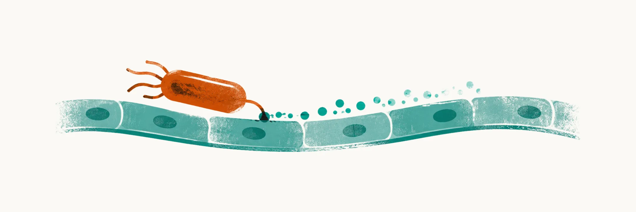
The urinary tract is a one-way line. The kidneys make urine, the ureters carry it to the bladder, and the urethra lets it out. The constant flow from kidney to bladder is the first thing that washes bacteria away. The uroepithelium is the surface where the contest between bacteria and the tract is decided.
A urinary tract infection (UTI) means bacteria are inside the urinary tract and the body has reacted to them. The reaction is the operative part. Inflammation in an infected kidney is what causes flank pain, and mediators carried in the blood spread the response beyond the kidney, so the symptoms come from the reaction more than from the bacteria alone. Bacteria can also sit in urine without inflammation or symptoms, and that state is not an infection.
In a child, three problems stack. First comes a bacterial adhesion problem: the bacteria must stick to the lining. Second comes an organ-defence problem: the tract must fail to clear them. Third comes an inflammatory problem, and this one can leave the kidney permanently scarred. If you hold these three layers together, the diagnosis, the imaging decisions and the long-term renal risk follow from the same mechanism.
So why do bacteria in the urine not always cause infection?
Why do bacteria in the urine not always cause infection?
Bacteriuria means bacteria are present in urine without an inflammatory response. Bacteria that enter the urethra do not automatically cause infection. Whether they do depends on how well the tract clears them, and that contest is decided at the uroepithelium.
Three defences matter, in descending order of importance. The first is unidirectional urine flow: the constant stream from kidney to bladder washes bacteria out. When flow slows, because of obstruction, stasis or incomplete emptying, bacteria have time to adhere to the lining.
Flow washes bacteria out, so the next question is what protects the surface they would stick to.
What else lines and protects the tract?
The second defence is an intact uroepithelium. It is a continuous layer of cells over the surface, so bacteria meet a barrier and cannot cross a continuous mucosal surface unless it is damaged.
The third is acidic urine pH. It helps, but it is not the main barrier.
Several secreted and surface factors support the lining:
- Uromodulin (Tamm-Horsfall protein) is secreted by cells of the thick ascending limb of the loop of Henle, so it enters the urine from the kidney. It binds type 1 fimbriae and traps bacteria in the lumen, so those fimbriae cannot bind the uroplakin receptors of the lining.
- Antimicrobial peptides are secreted into the urine.
- A glycosaminoglycan layer coats the umbrella cells of the bladder lining and resists bacterial binding, so bacteria meet a coated surface.
Flow and an intact surface carry most of the weight. The defence hierarchy is urine flow, then intact uroepithelium, then acidic pH as an assistant, not the main barrier.
Which bacteria get past these defences?
The organism that causes most infections is the one best equipped to hold on against the flow. E. coli that cause UTI are specialised strains adapted to live in the urinary tract.
In classic teaching, E. coli causes about 70–80% of childhood UTIs. A recent tertiary-center series found E. coli in 64%. Klebsiella, Pseudomonas, Enterobacter, Proteus and Enterococcus make up most of the remainder. Viruses rarely cause primary UTI.
E. coli is the organism that holds on, so how does it do so?
How does E. coli hold on?
E. coli carries fimbriae. These protein filaments bind uroepithelial receptors and let the bacterium hold its position against urine flow.
Type 1 fimbriae bind mannose-containing receptors. The mannose chains on Tamm-Horsfall glycoprotein change how well they bind. P fimbriae bind glycolipids on renal pelvic epithelium, and they act with type 1 fimbriae to colonise the kidney.
Adhesion is what allows ascent from the bladder to the kidney. Without it, the upper tract is difficult to reach.
What about organisms that do not hold on well?
Klebsiella, Pseudomonas, Proteus and Enterococcus do not share E. coli’s adhesin toolkit. Adhesins are what let E. coli colonise the kidney, so organisms without that toolkit climb the tract poorly on their own.
Ascent is only one of the routes bacteria use into the urinary tract. When these organisms appear in a young child’s urine, they more often arrived through a urinary tract that carries urine (and bacteria) upward.
A single-center series of 139 children hospitalised with a UTI is consistent with this. Of 32 children infected by a non-E. coli organism, 14 (43.8%) had an underlying urinary tract malformation. Of 107 children infected by E. coli, 4 had one. The difference between these groups is p < 0.0001. Looked at the other way, 14 of 18 children with a malformation were infected by a non-E. coli organism.
E. coli has adhesins that the other organisms lack, so what does their appearance in a young child mean?
What does a non-E. coli infection change?
A febrile UTI in a young child caused by a non-E. coli organism raises the probability of an underlying structural abnormality. An upward route provided by a structural abnormality is consistent with such an organism reaching the kidney. So this finding points towards a structural abnormality, not just towards a different antibiotic.
The rule shifts the workup towards imaging, particularly cystography. Cystography is an X-ray study that follows contrast in the bladder as the child voids.
An organism that does not climb well points away from the usual route. What, then, happens when the usual route does work and the bacteria arrive?
What happens once the bacteria arrive?
Adhesion explains how E. coli reaches the kidney. The illness comes from the host’s response once it arrives.
Three sequential events produce the illness. The first is adhesion: fimbriated E. coli reaches the renal pelvic uroepithelium and binds it. Without binding, the kidney is safe. The other two events are the lining’s recognition of the bacteria and the cytokine response that follows.
Arrival alone does not make the illness, so how does the lining sense the bacteria?
How does the lining detect E. coli?
Lipopolysaccharide (LPS) is a component of the outer wall of E. coli. It engages the TLR4/CD14 complex on the uroepithelial cell surface. TLR4 is the recognition molecule for LPS, and it works with its co-receptor CD14 on epithelial cells. On uroepithelial cells, membrane-anchored CD14 is important for LPS to induce IL-8.
TLR4 signalling runs through an intracellular cascade that activates NF-κB. This transcription factor translocates to the nucleus.
Why does the child get a fever?
The lining’s signal reaches the body as cytokines. NF-κB drives production of IL-6, which is thought to act on the hypothalamic set point and so produce the fever. The evidence for this is from animal studies, so the mechanism is proposed rather than proven.
The fever is therefore a cytokine event, not a direct effect of bacteria on the brain. That is why the temperature can be high while urinary findings look modest, and why a child can be hot with only modest urinary findings.
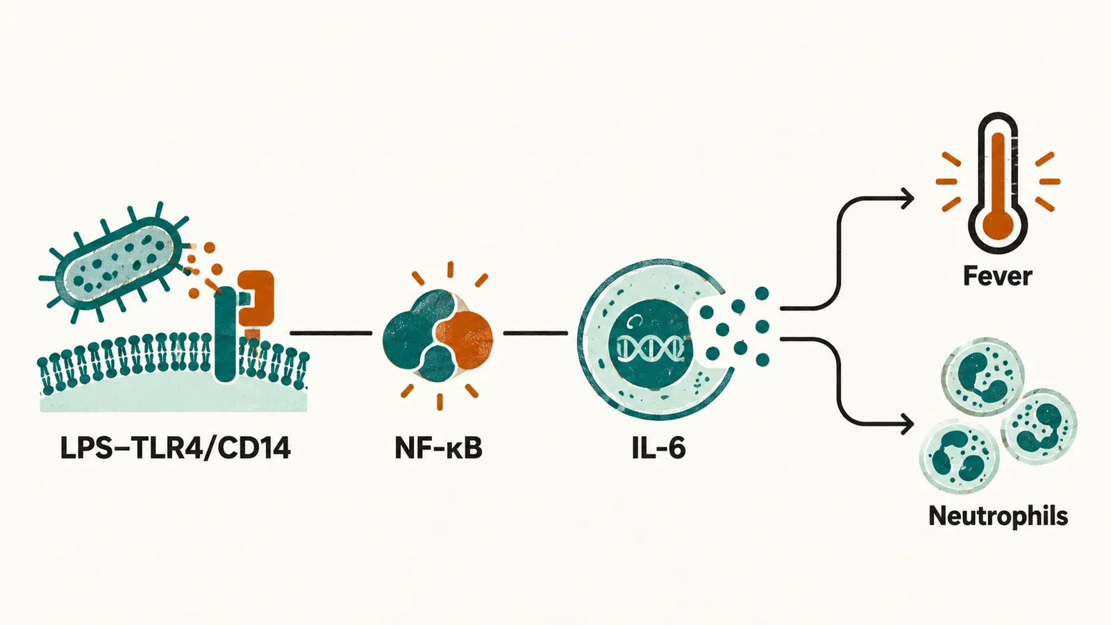
The inflammatory chain runs: fimbrial adhesion, then LPS–TLR4/CD14, then NF-κB, then IL-6 (fever), TNFα and IL-8 (neutrophils).
What brings the white cells into the urine?
The same cytokines call neutrophils. TNFα and IL-8 increase local vascular permeability and recruit neutrophils. Neutrophils are responsible for pyuria (white cells in the urine) and for the inflammatory infiltrate of pyelonephritis.
Does fever mean the kidney is involved?
Fever comes from mediators that have spread from the infected kidney, so fever is a sign that the response has reached beyond the bladder. Febrile UTI is infection that has reached the renal parenchyma, with fever as its signature. A febrile UTI in a child means renal involvement until shown otherwise.
In pediatric practice, a febrile UTI is treated as pyelonephritis. Pyelonephritis means infection of the renal parenchyma (the functioning tissue of the kidney), not bladder-limited disease. Most febrile infants and young children with a UTI show acute renal parenchymal changes on scintigraphy (nuclear scan of the kidney).
Fever points to the kidney, so where is the clinical boundary drawn?
Where is the line between bladder and kidney infection?
Cystitis is inflammation confined to the bladder, usually without fever. Fever and flank pain come from inflammation in the kidney, so a syndrome limited to the bladder does not carry them. An “afebrile UTI” therefore does not by definition mean the kidney is uninvolved. It means the clinical syndrome is limited to the bladder.
Guidelines draw the boundary between upper and lower UTI clinically rather than radiologically. Bacteriuria with a temperature of 38°C or higher, or bacteriuria with a temperature below 38°C plus loin pain or tenderness, is treated as acute upper UTI (pyelonephritis). Bacteriuria without systemic symptoms or signs is lower UTI, or cystitis.
The temperature is a threshold for action, not a measure of how much kidney is involved. A temperature of 38°C or higher is the clinical boundary of upper tract disease.
Does an acute lesion always leave a scar?
A febrile kidney infection is an acute lesion of the renal parenchyma. Experimental pyelonephritis links the inflammation itself to the scarring that follows, so scarring is the lasting trace of the inflammatory response, and the inflammatory problem can leave the kidney permanently scarred. But scarring follows a febrile infection in only a subset of patients, so an acute lesion does not always become a permanent scar.
How often does a first febrile UTI happen at each age, and which children does it affect?