The hardest part of diagnosing a urinary tract infection in a child is not choosing the antibiotic. It is obtaining a specimen that means something. Young children do not void on command, they cannot aim, and they cannot discard the first part of the stream. That is why the sample needs a sequence.
The urine sample depends on the route it took. The bladder is what the sample should reflect, but in a child the urine can pass over the skin around the genitals, and that skin carries its own bacteria. The two strip tests read urine indirectly: one reads white cells the body has sent in, the other reads a chemical that some bacteria make, and neither counts organisms. The culture counts organisms, so it needs a sample that has not picked up skin flora, and it must be taken before an antibiotic changes what grows. Each step answers one question: which sample, which test, and how many colonies count.
Why is the sample collected in stages?
Most services use a tiered sequence. A non-sterile screening specimen decides whether a sterile specimen is needed at all. The first stage is a bag, and the bag picks up skin flora, so the sequence exists to separate skin from bladder.
The first step is a bag specimen: a sterile collection bag is applied to the perineum after local cleaning, and it is removed as soon as the child voids. The skin around the genitals is colonised by commensal bacteria, and a bag stuck to it gathers them along with the urine. So the bag is a screen and not a culture sample.
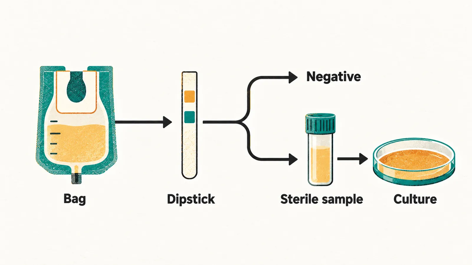
Why can the screen tolerate this, when a culture cannot? A culture counts each organism that grows, and it must grow a single organism from a specimen that reflects the bladder. Skin flora growing alongside the bladder’s organism spoil the count. So the culture needs a second-stage collection. The order is therefore bag, then dipstick, then culture, and the culture is sent only when the dipstick supports it.
What does the dipstick read?
The second step is the dipstick, run on the bag urine. It reads leukocyte esterase and nitrite. The dipstick tolerates perineal flora because it detects the body’s response rather than the organism. The dipstick reads the bag sample, so what does it actually measure?
What a positive leukocyte esterase means
The first reading, leukocyte esterase, tells you about the body’s response. Leukocyte esterase is a proxy for white cells in the urine, meaning the bladder is inflamed enough to recruit neutrophils. The strip detects the white cells that have reached the urine and not the bacteria themselves. The white cells the inflamed bladder draws in are neutrophils, as the earlier foundations note, Pediatric UTI: Foundations and Pathophysiology, explains. Leukocyte esterase reports the host response, not the bacterium.
What the nitrite reading means
The second reading comes from bacteria, not from the body, and that changes what a negative means. Nitrite is produced when Gram-negative bacteria reduce dietary nitrate in the urine. So a positive nitrite points to bacteria. It is relatively specific for E. coli, Klebsiella and Proteus, and a positive result is seldom false.
A negative result is different. Not every E. coli strain reduces nitrate efficiently, so a negative nitrite does not exclude E. coli. Enterococcus and Pseudomonas frequently do not reduce nitrate at all, so nitrite may not signal their infections. In short, a positive nitrite is specific, and a negative nitrite is not sensitive: infections can still give a negative result.
Why the urine must be read quickly
Nitrite is formed in the sample itself, so timing matters. A sample left at room temperature can become falsely positive as the bacteria keep multiplying. The urine should therefore be examined promptly.
How much can the pair tell you?
The question is how well the two readings work together. The combined dipstick performs best as a pair. Leukocyte esterase reports the body’s response and nitrite reports the organism, so the two can catch different infections.
Performance is expressed as a likelihood ratio, and values well above 1 raise the odds of UTI, while values below 1 lower them. A dipstick positive for both leukocyte esterase and nitrite has a pooled positive likelihood ratio of about 28 for UTI. A dipstick negative for both has a pooled negative likelihood ratio of about 0.2. Even so, a normal urinalysis does not exclude infection. Automatic urinalysis is reported to miss a substantial minority of UTIs in febrile infants and toddlers when pyuria is absent.
What about results that do not fit?
Some children fall between the results, and that is where judgement enters. Children with a positive urine culture but no pyuria sit in an ambiguous zone. Some of them have asymptomatic bacteriuria or a contaminated specimen. Bacteria can sit in urine without any inflammatory response, as the earlier foundations note explains. Others have a genuine infection with a falsely negative screen. Therefore the clinical condition, the collection method and the decision to treat all enter the final judgement, not the dipstick alone.
What is done with each result?
A negative screen and a positive screen lead to different next steps. If the dipstick is negative, do not culture: the probability of UTI is low enough that a culture adds little. If the dipstick is positive, obtain a sterile specimen and send it for culture. A positive dipstick always needs confirmation with a sterile sample, because bedside dipstick tests are useful for screening but urine culture is required for diagnostic confirmation.
The practical rule is asymmetric. A negative dipstick in a well child can reasonably end the workup. A negative or equivocal dipstick in an unwell febrile child should not by itself stop a culture being sent, because a normal urinalysis does not exclude UTI. The estimated probability of infection decides whether a specimen is sent at all, as the earlier epidemiology note, Pediatric UTI: Epidemiology and Pre-Test Probability, shows.
How is a sterile sample collected?
A positive screen needs a sterile sample, and three methods can supply one. Whenever a culture is needed, the specimen has to reflect the bladder rather than the skin, so a specimen taken from the bladder avoids the skin route. The choice depends on whether the child can cooperate and how unwell the child is.
- Clean-catch midstream: used for a cooperative, toilet-trained child. The perineum is cleaned, the first part of the stream is discarded to clear urethral flora, and the middle is caught.
- Catheter specimen: used for an unwell child, suspected pyelonephritis, or when an urgent sample is needed. It is invasive and uncomfortable, and it must be aseptic, since a catheter can introduce organisms into the bladder.
- Suprapubic aspiration: rarely used, when catheterisation is not feasible or has failed. A needle aspirates the bladder through the abdominal wall, so the urine does not pass the perineal skin, which is why it avoids perineal flora.
Collecting from an infant who cannot void on command
The least invasive method for an infant depends on stimulating voiding. For precontinent infants, bladder stimulation methods can help an infant who cannot void on command give a clean-catch sample. They are gentle suprapubic tapping and lumbar stimulation, or the Quick-Wee variation, which uses gauze soaked in cold fluid applied to the suprapubic area. In a randomised trial, bladder stimulation increased the proportion of infants who voided within 5 minutes compared with an unaided clean-catch attempt. This non-invasive route is therefore more often successful. Published success rates vary widely between studies and depend on the operator’s technique.
How many colonies count as infection?
A sterile sample is cultured, and the colony count has to be read against the method used. Results are reported in colony-forming units per millilitre (CFU/mL): the count of bacteria growing from each millilitre of urine. Each collection method lets in a different amount of skin flora, so the threshold for a significant count differs by method.
- Clean-catch or bag: usually significant at ≥ 100,000 CFU/mL (10⁵) of a single organism.
- Catheter: usually significant at ≥ 10,000 CFU/mL (10⁴), and often any growth of a single organism.
- Suprapubic aspirate: usually significant at any growth of a single organism (≥ 10³).
So the thresholds are ≥ 10⁵ CFU/mL for clean-catch, ≥ 10⁴ CFU/mL for catheter, and any growth from a suprapubic aspirate.
Why dilute urine can still count
Concentration changes what the count means. In very dilute urine, with a low specific gravity, lower counts of 60,000–80,000 CFU/mL of a single organism can still be significant, because the measured concentration is diluted by the larger volume.
Pyuria, the count and contamination
The count is read together with the urinalysis. Pyuria plus a significant single-organism count supports the diagnosis. Mixed growth usually indicates contamination, which is what skin flora produce.
Which guideline sets the cut-off?
Thresholds differ between guidelines, so the local definition decides. The AAP guideline for febrile infants and children aged 2–24 months defines UTI as pyuria together with at least 50,000 CFU/mL of a single uropathogen. Other guidelines set different thresholds, and the definition in use locally should be the one applied.
Why is the sample taken before the antibiotic?
A culture is less useful if an antibiotic has already suppressed the bacteria it would grow. Antibiotics suppress bacterial growth, so a specimen taken after treatment has started frequently comes back sterile or with an uninterpretable result. The order is fixed: collect the specimen first, then give the antibiotic, even in a child who looks unwell. Specimen first, antibiotic second, in every child.
What the child needs while the culture grows
The child needs both treatment and diagnosis. Culture takes 24–48 hours, and a child with febrile UTI is at risk of bacteraemia and urosepsis, so treatment still starts before the culture result is available. The sequence is the specimen, then an empiric antibiotic, then adjustment once the organism and its susceptibilities are known. Which antibiotic should be started while the culture grows, and does the site of the infection decide that choice?