
During a febrile UTI the renal parenchyma is inflamed. In most children the inflammation resolves with treatment and leaves nothing behind. In a minority it ends as a renal scar. What is a scar, and how is it seen?
What does a scar look like on the scan?
A kidney filters blood through its nephrons. A renal scar is a focal area of cortical loss where functioning tubules have been replaced by fibrous tissue. Where inflammation destroys tissue rather than only swelling it, the gap heals with fibrous tissue, which does not filter. The tubular cells in that area have stopped regenerating, so the scar is fibrous replacement of nephrons already lost.
A scar is found with a DMSA scan, in which the tracer is taken up by functioning tubular cells. On the scan a scar shows as a patch with no tracer uptake. As the imaging note set out, DMSA is a nuclear scan that reads functioning kidney tissue, and a reduced uptake marks scar but can also come from the acute infection.
A scan during the acute illness shows which parts of the kidney are involved in the infection. The defect seen then may disappear as the inflammation settles. As the foundations note set out, an acute lesion does not automatically become a scar. So a defect seen only during the acute illness is not a scar.
When is a defect a permanent scar?
A defect seen in the acute illness may be only inflammation, so the question is how long it lasts. The scan that identifies permanent damage is done 4–6 months after the infection, once healing is complete. A delayed DMSA scan at 4–6 months defines a scar.
A defect still present after healing is one where tissue was replaced, not only swollen, because arrested tubular cells do not regenerate. For the same reason, more than 90% of defects still visible at least 5 months after the episode persist: the lost tissue does not come back. More than 90% of defects present at 5 months persist. A scar found on the late scan is a scar for life, because the tissue it replaces does not regenerate.
How often does one febrile infection leave a scar?
Once a scar is defined, the next question is how often one first infection leaves one. After a first UTI, the pooled estimate of renal scarring on delayed DMSA is about 15% (95% CI 11–18%). This is the estimate from a systematic review of children imaged after a first UTI, not all of them strictly febrile. About 15% of children scar after a first UTI. Put the other way, approximately 85% of children recover without scarring after a first infection.
Why does a second infection matter more?
The rate after one episode is low, so what changes with a second? First, what counts as recurrence. As the foundations note set out, a febrile UTI is infection that has reached the kidney, and cystitis is infection confined to the bladder. Acute upper UTI is pyelonephritis, and lower UTI is cystitis. Recurrent UTI means two or more episodes of acute upper UTI, or one upper UTI with at least one lower UTI, or three or more lower UTIs.
The figures for febrile infections come from an analysis of 345 children, all of whose infections were febrile:
- After one febrile UTI: 2.8% scarring.
- After two febrile UTIs: 25.7% scarring.
- After three or more febrile UTIs: 28.6% scarring.
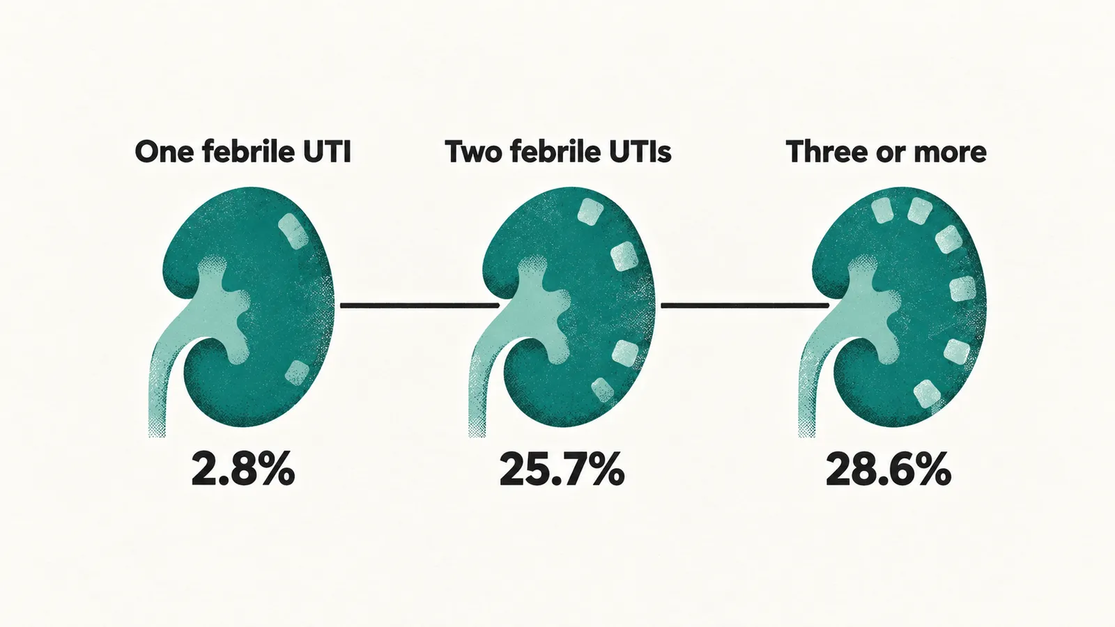
In that analysis the odds of scarring after a second febrile infection were about 12 times higher than after a single one. So recurrence, not the first episode, drives scarring. The reason is that each febrile infection that reaches the kidney is another round of inflammatory damage, and each round that leaves a scar takes away filtering tissue, so the losses add up. The risk rises sharply with each further febrile episode. The scarring risk is 2.8% after one febrile episode, 25.7% after two and 28.6% after three or more, and recurrence is the driver of scarring.
What does each scar cost the kidney?
Recurrence matters because each scar takes filtering tissue, so what does the kidney do about lost tissue? As the CAKUT note set out, the kidney starts life with a set number of nephrons, and when some are lost the survivors filter more so that total filtration holds.
The kidney starts with on the order of 1 million nephrons, with wide individual variation. After a segment is lost, the surviving nephrons compensate by hyperfiltrating: they enlarge and filter more of the blood, so total filtration holds. That is why one scar is usually absorbed without a measurable fall in GFR. A single small scar usually leaves kidney function normal, because the survivors compensate.
The mechanism behind the recurrence effect is nephron reserve. Repeated scars exhaust the reserve. When the survivors can no longer keep up, their strain raises pressure in the filtering units, and this shows as GFR declining and hypertension or proteinuria appearing.
Which children are most at risk?
Recurrence is not the only difference between children, so which findings mark those at highest risk? A pooled analysis of individual patient data covered 1,280 children with a first UTI, all scanned at least 5 months later. It identified findings that predict scarring, and the results are expressed as odds ratios. The strongest predictors are grade IV–V reflux, an abnormal ultrasound, and a temperature of at least 39°C combined with an organism other than E. coli.
Reflux grade
The strongest marker is reflux, so how does grade matter? As the reflux note set out, vesicoureteral reflux is backflow of urine from the bladder towards the kidney, and the grade reads how far contrast climbs the collecting system and how much it stretches it.
Scarring has a strong grade dependence. Grade IV or V reflux is the strongest predictor: the odds of scarring are about 22 times those of children without reflux, and roughly two-thirds of children with grade IV or V reflux scarred. Grade III reflux brings about a 3.6-fold increase in odds. Grades I and II are only marginally above no reflux. High-grade reflux is uncommon, present in about 4% of children.
Other markers
Beyond reflux, other findings add to the odds. An abnormal ultrasound (dilatation, abnormal parenchyma or a bladder abnormality) raised the odds about 3.8-fold. A temperature of at least 39°C had an odds ratio of about 2.3, and so did an organism other than E. coli. A C-reactive protein above 40 mg/L had an odds ratio of about 3.0, and a polymorphonuclear cell count above 60% had one of about 1.9. These last markers show a more intense infection, and markers of more intense infection also counted as predictors.
A child with an abnormal ultrasound, or with both a temperature of at least 39°C and a non-E. coli organism, has roughly twice the baseline risk of scarring. About one child in three in that group scars.
Age and sex
The markers one might expect from age and sex do not predict scarring, so why is that notable? Age, sex and the duration of fever before presentation were not associated with scarring. Older age and female sex were, if anything, weakly predictive. This runs against older studies, which identified the youngest children and boys as at greatest risk. Age and sex do not predict scarring.
Do most children who scar do well?
Given the risk, what usually happens to the kidney afterwards? Most children who scar do well. A review of the long-term evidence found that children with normal kidney function and renal imaging at baseline rarely lose kidney function over years of follow-up. The risk of hypertension is low and largely confined to those who already had renal damage. This fits the picture, because hypertension follows hyperfiltration, and hyperfiltration is what damaged kidneys do, so it is seen where there is damage.
Why does blood pressure give the first sign?
If most do well, what would show that the reserve is being spent? Blood pressure is the earliest measurable signal. Hyperfiltration raises pressure in the filtering units first, and systemic hypertension follows, so pressure is the earliest measurable change. Children with higher grades of scarring show day- and night-time blood pressure elevation, because once systemic hypertension follows hyperfiltration the raised pressure is present at all times of day.
Why does pregnancy matter?
Pregnancy is the other occasion on which reduced renal reserve becomes visible. Pregnancy raises the demand for filtration. A kidney with little reserve cannot meet it, although a kidney with intact reserve usually can. Controlled cohorts have not found an overall increase in first-pregnancy complications among women with childhood UTI. The pregnancy risks are concentrated in women whose kidneys carried measurable damage.
How are these children followed?
Blood pressure and kidney function are what is watched, so who watches them, and how is the watching matched to the damage? Follow-up is matched to the amount of damage rather than offered uniformly. Two findings reliably trigger specialist assessment: abnormal imaging, and recurrent UTI.
A child with a renal parenchymal defect is assessed with height, weight, blood pressure and routine testing for proteinuria. Blood pressure, urine protein and kidney function are the readings that show the filtering units under strain. Minor unilateral defects need no long-term follow-up unless there is recurrent UTI, a family history of hypertension, or lifestyle risk factors for hypertension.
Bilateral abnormalities, impaired kidney function, raised blood pressure or proteinuria warrant monitoring and management by a paediatric nephrologist. The aim of that management is slowing the progression of chronic kidney disease. Blood pressure, proteinuria and kidney function are monitored closely where damage is bilateral or function is already reduced.
Urine is not retested routinely in an asymptomatic child after an episode. Asymptomatic bacteriuria is not a reason for follow-up.
Was all of it scarring?
Some of what was called post-infectious scarring was never caused by infection, which changes what the follow-up is for. As the CAKUT note set out, renal dysplasia is disorganised kidney tissue that formed abnormally before birth. Part of what was called post-infectious scarring turned out to be congenital dysplasia, visible on antenatal ultrasound. The old assumption that childhood UTI is a common route to kidney failure is now debated. The proportion of end-stage renal disease attributable to childhood UTI appears small.
What does the long view show?
Where damage is established from the start, the outlook is less benign. What do the long series show?
One is a 27-year follow-up of 30 adults with non-obstructive focal scarring after childhood pyelonephritis. In it, 7 developed hypertension and 3 reached end-stage renal disease. Two of 16 women had a history of toxaemia in pregnancy. Even patients who escaped surgery or kidney failure had a lower GFR than age-matched controls. The series is small, selected and decades old, so it describes the severe end of the spectrum rather than the average outcome.
What stops the next scar?
The long view shows that outcome depends on what is left, so what stops the next episode from adding damage? Prevention is aimed at the second and third febrile UTI rather than the first, which reveals the underlying substrate. Any suspicion of recurrence is assessed promptly, since the next febrile episode adds to the damage.
Constipation and dysfunctional elimination are treated. As the bladder and bowel note set out, urine left in a bladder that empties incompletely keeps the infection going. Children are encouraged to drink enough to avoid dehydration, and are given access to clean toilets without having to postpone voiding until unavoidable.
Antibiotic prophylaxis is not routinely given after a first UTI. It is not given for asymptomatic bacteriuria either. Where reflux is present, the decision is different and more finely balanced. Prophylaxis reduces recurrent infection but has not been shown to reduce new scarring. It is reserved for selected higher-risk children. In short, prevention targets the next febrile episode: bowel and bladder management, adequate fluids, prompt voiding, and selective prophylaxis in reflux.
What is still not known?
The prevention question leaves two matters open: which children belong to the minority who scar, and what a scar means over the following decades.