
How does a normal gland come to fail later in life?
Acquired hypopituitarism develops after birth, when damage to the pituitary or to the hypothalamus above it stops the gland from making its hormones. The pituitary makes its hormones only when told to, and the hypothalamus sends its signals down the stalk, so a break at the stalk or in the gland lowers hormone output.
The causes differ in how they reach the gland. A normal gland can be damaged by a small number of routes:
- compression from around the sella, the bony hollow at the skull base that holds the pituitary
- an interrupted blood supply
- infiltration or inflammation of the gland
- direct injury, as trauma does
- irradiation
Infection of the region also belongs here, and it is covered with the infiltrative diseases. The routes differ in tempo. A bleed is sudden, while an autoimmune attack, an infiltrating disease or a tumour is slow, and irradiation is slower still.
Which tumours cause it?
The sella is a bony cup with no room to expand, so a growing mass presses on the gland. A pituitary adenoma is the most common cause of adult-onset hypopituitarism. It does this whether or not it secretes a hormone: a non-secreting adenoma still occupies space, so what matters is the mass, not its hormone.
Craniopharyngioma is the next most prevalent tumour in this group. It lies near the gland and the stalk and is thought to arise from embryonic remnants. It is more common in children, with a peak age of 10-20 years. Rathke’s cleft cyst is more often found in adults. Metastatic tumours and lymphoma are further neoplastic causes of hypothalamic-pituitary damage.
A mass can do more than press. It can also outgrow its blood supply.
What happens when a tumour or a pregnant gland suddenly bleeds or dies?
Pituitary apoplexy is the clinical consequence of haemorrhage or infarction in a pre-existing pituitary adenoma. A growing tumour can have blood-flow demands that exceed its supply, so it can die from lack of blood, and it can also bleed within itself. Apoplexy can occur in several settings. It may follow a known adenoma, or it may be the first event.
Apoplexy can also occur after a postpartum event, known as Sheehan’s syndrome. Here the cause is hyperplasia of the pituitary during pregnancy, which raises the risk of haemorrhage and infarction. An enlarged gland has been suggested to be more exposed when it loses blood supply after delivery.
Predisposing factors for apoplexy are hypertension, diabetes, sickle cell anaemia and acute shock. In hypertension, an acute increase in pituitary blood flow can precipitate a bleed; in shock, low blood supply can give pituitary ischaemia. Hypertension was found in 26% of patients with apoplexy.
Apoplexy is an endocrine emergency, because the whole gland can fail at once. Two things happen together. First, blood or swollen tissue raises the pressure inside the sella, and this pressure has to go somewhere. That explains the picture:
- sudden severe headache
- visual disturbances, because the optic chiasm lies above the gland and is compressed from below
- ophthalmoplegia from damage to cranial nerve III, IV or VI, because these nerves run beside the gland, through the cavernous sinus, and are affected by lateral compression
Second, the hormones are lost at once. ACTH tells the adrenal glands to make cortisol, so losing ACTH means losing cortisol. Cortisol keeps the vessels responsive to catecholamines, and without it blood pressure falls. This fits the other manifestations of apoplexy: severe hypoglycaemia, hypotension and shock.
Finally, apoplexy can manifest as CNS haemorrhage, since blood can escape beyond the sella into the subarachnoid space.
Can the vessels beside the sella cause it?
A tumour is not the only mass that presses on the gland. An aneurysm near the sellar region can compress the gland and cause hypopituitarism, by the same pressure route. Such an aneurysm is most often of the suprasellar or infrasellar carotid arteries, or a suprasellar aneurysm of the anterior or posterior communicating arteries.
How can inflammation do it?
After pressure and blood supply, the immune system can be the route. Lymphocytic hypophysitis is autoimmune inflammation of the gland, in which overly active lymphocytes damage tissue. It mostly occurs in postpartum women, so it arises at the same time as Sheehan’s syndrome. It is now thought that a significant number of patients previously diagnosed with Sheehan’s syndrome actually had lymphocytic hypophysitis.
Prolactin offers a clue. Lymphocytic hypophysitis produces hyperprolactinaemia, because the inflammatory process interrupts dopamine inhibition of the lactotroph cells. Prolactin therefore rises while other hormones fall. MRI shows a prominent pituitary mass that resembles an adenoma, so imaging alone can mislead.
A lineage is one hormone-producing cell type, so damage can hit one or several. Hypopituitarism in lymphocytic hypophysitis usually affects all cell lineages. In rare cases it is isolated. The mass also compresses its neighbours, which adds visual disturbances and headache, the same pressure picture as in apoplexy.
What about diseases that infiltrate and infections?
Inflammation is one way to fill the gland; infiltration is another. Infiltrative disease can involve both the hypothalamus and the pituitary. It is most often seen in:
- sarcoidosis
- amyloidosis
- histiocytosis X
In more than half of these patients, diabetes insipidus is part of the picture. Growth retardation appears if the disorder occurs before puberty and interferes with GH release. Hyperprolactinaemia and hypogonadism are also common in infiltrative disease.
Infection of the region is uncommon but recognised. The usual settings are:
- fungal infection after AIDS
- bacterial infection with tuberculosis
- tertiary syphilis
Can a cancer drug cause it?
The immune attack can also be drug-induced. Pituitary cells express CTLA-4 extensively, and immune checkpoint inhibitors that block CTLA-4 can produce hypophysitis. A proposed explanation is that the blocking antibody binds CTLA-4 on pituitary cells and triggers an immune attack on them; this is thought to be how the drug acts, not an established cause.
Checkpoint-inhibitor hypophysitis is pituitary inflammation like the autoimmune form. Involvement is heterogeneous, and which cell types are lost varies with the type of inhibitor. It may extend to the adrenal gland, gonadal activity and the thyroid gland.
How does trauma damage the gland, and how often?
So far the gland was harmed from within. Trauma harms the pituitary or its stalk from outside. The gland is held in the cup and tethered to the hypothalamus by the stalk, so impact, skull-base fracture and shearing can injure the gland or the stalk, and bleeding or low blood flow after major trauma can add ischaemic injury. The long portal vessels of the stalk run across the diaphragma sellae and are thought to be particularly vulnerable.
Studies show that about 33% of traumatic brain injuries have hypopituitarism 3 months after trauma. After 12 months of follow-up, 22% are still affected. The drop from 33% to 22% hints that part of the early loss recovers. Neurosurgical trauma to the region produces the same kind of injury.
Why does radiation damage appear years later?
Radiation is also an external injury, but it works slowly. Cranial irradiation can damage the hypothalamus and the pituitary, and the site of damage has been debated, with evidence for direct pituitary injury and for hypothalamic injury. The damage is greater in children and adolescents.
The degree of hormone deficiency depends mainly on the irradiation dose and the time since completion. About two-thirds of patients receiving at least 50 Gy to the skull base develop a hormone deficiency. The damage is progressive and develops over time, so deficiency usually appears 5-15 years after radiotherapy, and a normal early test does not exclude later loss. The usual order of loss is:
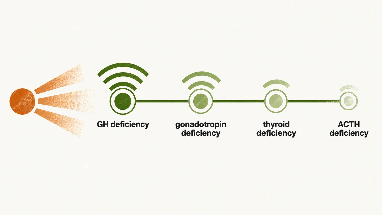
- GH deficiency
- gonadotropin deficiency
- thyroid deficiency
- ACTH deficiency
Why follow patients whose deficiency looks transient?
After trauma, surgery or irradiation the picture can change over time. Post-traumatic, post-surgical or post-irradiation hypopituitarism may be transient and short-lived, because some of the early loss is reversible. Long-term follow-up is still indicated in these cases, because in about 25-40% of these patients the condition progresses to long-term hypothalamic or pituitary dysfunction.
Whichever way the gland was damaged, how does a patient with missing hormones actually present? See Clinical manifestations of hypopituitarism.