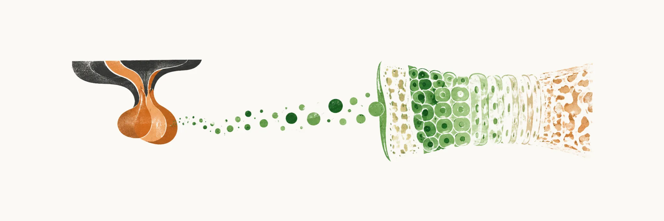
How does GH make a child grow?
Growth runs on a short chain. The anterior pituitary makes GH, and GH acts partly through IGF-1, which cells produce in response to GH. That is why GH deficiency presents differently by age: a child still has open growth plates, and an adult does not.
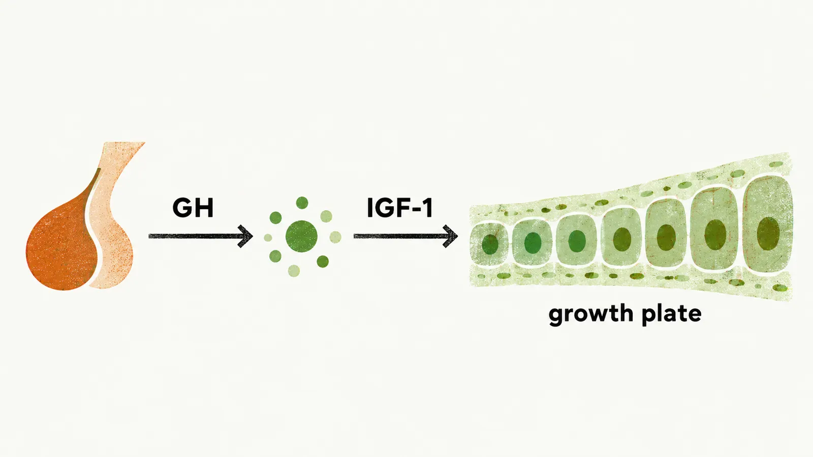
Growth is fastest in infancy, when it depends mainly on the pituitary gland. After infancy the average growth rate is about 6 cm per year, with another growth peak in mid-puberty, at age 12 in girls and 13 in boys.
The end of growth is set at the growth plate, and closure of the plate depends on several endocrine factors, including GH, IGF-1, sex steroids and TSH. Paracrine growth factors, such as FGFs, are also involved. Other influences act at the same level: energy expenditure, nutrition, vitamins and metals.
What else slows growth, besides the hormone?
If IGF-1 carries much of the GH effect, anything that lowers its action slows growth. Malnutrition has its own endocrine effects, and it does so by three routes:
- It causes resistance to GH at the chondrocytes, so the growth plate responds less to the hormone.
- It causes a reduced IGF-1 level.
- It causes reduced production of IGFBP-3 (IGF-1 binding protein-3), the protein that carries IGF-1 in the blood.
How does GH deficiency look in a child?
In children the main sign is slowed growth. The GH and IGF-1 chain feeds the growth plate, so when GH is missing, the plate is not driven to grow.
In all true forms of GH deficiency or GH receptor dysfunction, bone age is delayed compared with the norm for age. Delayed bone age with short stature in a child therefore suggests GH deficiency. Short stature here means a height at the 3rd percentile.
Isolated GH deficiency in childhood has other features too: micropenis, increased fat tissue, which is attributed to the lipolytic effect of GH being lost, and a high-pitched voice. The child also has a higher chance of hypoglycaemia, because there is less of a hormone force opposing insulin.
Why does a child lack GH?
Lacking GH means that something along the route to it is faulty: the gland itself, the receptor that hears the hypothalamic signal, or the transcription factors that build the hormone-making cells. Familial inherited forms, autosomal dominant or X-linked, account for about 33% of childhood GH deficiency cases.
Mutations in the transcription factors Pit-1 and Prop-1 cause combined, syndromic deficiency, not an isolated loss of GH, because these factors are needed for several hormone cell types. A recessive mutation in the GHRH receptor causes severe dwarfism: the signal from the hypothalamus is not heard, so the somatotropes make little GH. It brings:
- a low basal GH level,
- increased susceptibility to insulin-induced hypoglycaemia,
- anterior pituitary hypoplasia.
Not every cause is a gene. Psychosocial deprivation is a cause of GH deficiency in children. Emotionally and socially deprived children can show:
- short stature,
- hyperphagia,
- delayed speech,
- an attenuated response to GH administration, since the child’s psychosocial environment is one of the factors that can blunt the response to GH.
When no cause is found, the deficiency is called idiopathic, and idiopathic GH deficiency is the remainder.
What if the hormone is there but the cells do not respond?
GH insensitivity is a different problem from GH deficiency: in deficiency the hormone is lacking, and in insensitivity its effect is lacking. In Laron syndrome, loss-of-function mutations of the GH receptor leave it partially or completely unresponsive. GH cannot signal through the receptor, so IGF-1, which is normally produced by cells in response to GH acting through that receptor, stays low. GH levels are normal or high, and the high GH fits with the missing negative feedback by IGF-1.
GH insensitivity can also be secondary. Each of these makes the tissues respond less to GH:
- malnutrition,
- uncontrolled diabetes,
- chronic renal failure.
How is GH deficiency tested in children?
GH secretion is pulsatile, so a single sample can fall between peaks and say little. The best way to assess GH level in children is therefore a provocative test: post-exercise, insulin-induced hypoglycaemia, or a pharmacological stimulus. A normal response raises GH above 7 microgram/L.
Before GH testing it is mandatory to ensure that thyroid and adrenal hormone levels are normal. Thyroid and adrenal hormones influence GH secretion and would confound the result.
What does GH deficiency do in an adult?
In an adult the growth plates are closed, so the same lost hormone now looks different: adults show changes in body composition and cardiovascular risk. Adult GH deficiency (AGHD) is mostly caused by acquired damage to hypothalamic or pituitary somatotrope cells, which are the cells that make GH. Acquired damage reaches a normal gland by compression, loss of blood supply, inflammation, infection and irradiation.
Acquired pituitary hormone deficiency normally follows a pattern, with GH failing first. The order of failure is GH, then FSH/LH, then TSH, then ACTH. Prolactin deficiency is very rare, and is seen mainly in Sheehan’s syndrome.
The main clinical manifestations of AGHD fall into three groups: body composition changes, cardiovascular effects and psychosocial effects.
Body composition and bone
GH normally has a lipolytic effect and supports lean mass, so with GH lacking, lean body mass decreases and fat tissue increases. The increase in fat is particular in intra-abdominal and visceral fat, and the waist-hip ratio rises. Bone deposition also decreases, so bone is laid down less and the risk of bone fracture is increased.
Cardiovascular effects
The cardiovascular findings are:
- reduced exercise tolerance, which may be explained by the fall in lean body mass and possibly by reduced left ventricular function,
- hyperlipidaemia, with increased LDL-cholesterol, total cholesterol and apolipoprotein B,
- left ventricular dysfunction, described in studies,
- hypertension, for which a proposed explanation is reduced nitric oxide synthase activity with higher peripheral vascular resistance,
- increased plasma fibrinogen.
Hypopituitarism carries a roughly two-fold increase in cardiovascular mortality. Unreplaced GH deficiency is a suggested important contributor to these excess deaths, and the increased cardiovascular mortality is probably mainly related to GH deficiency. The adverse lipid, fat, vessel and pressure changes are the kind that would predict more atherosclerosis.
Psychosocial effects
AGHD impairs quality of life and general well-being. How GH deficiency produces this is not well understood.
Who should be tested as an adult, and how?
The adult picture is subtle: the symptoms are common, and essentially no feature is specific to AGHD. Testing therefore follows the settings where hypothalamic-pituitary damage is likely. Testing for AGHD is recommended only in patients with:
- pituitary surgery,
- pituitary or hypothalamic tumours or granuloma,
- a history of cranial irradiation,
- radiological evidence of pituitary lesions,
- a childhood requirement for GH administration.
It is not true that a patient who was GH deficient in childhood or adolescence is still GH deficient in adulthood, possibly because the hypothalamo-somatotroph axis matures. Retesting is always needed. In children with idiopathic isolated GH deficiency, about three-quarters no longer meet the criteria when retested at final height.
IGF-1 cannot settle the question. It behaves the same way in adults and in children: in a significant proportion of patients with true GH deficiency, IGF-1 lies in the lower normal range, so a normal value does not exclude it. IGF-1 is not an index for diagnosis. It is used for evaluation of treatment.
The most validated test for AGHD diagnosis is GH measurement after insulin-induced hypoglycaemia, a potent stimulus of GH release. AGHD is defined as a peak GH level below 3 microgram/L after the provoking test.
How sure is the diagnosis in each group of adults?
Adults with GH deficiency fall into three groups:
- Prior childhood GH deficiency.
- Adult-onset GH deficiency acquired through structural damage.
- A rare idiopathic form.
The groups differ in how much supports the result. In the idiopathic form there is no pituitary lesion or other hormone deficiency to back the test, so the diagnostic criteria are stricter: at least two positive dynamic tests are needed.
GH usually fails first, and the other axes fail after it in the order given, with the ACTH and gonadotropin axes among them. So what does the patient lose when those two axes go?