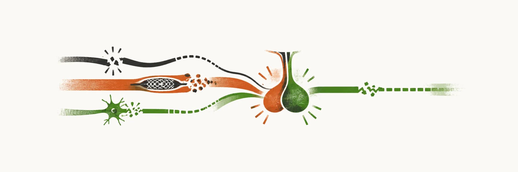
What can go wrong between the hypothalamus and the hormone?
Hypopituitarism is deficient production of one or more pituitary hormones. The pituitary is a small relay at the base of the brain, and its hormones drive the thyroid, the adrenal glands, the gonads and growth. The gland makes a hormone only when it is told to. The hypothalamus above it sends releasing signals down the stalk, and each cell type in the anterior gland answers by making its own hormone.
So there is a chain: the hypothalamus, the stalk, the cell type, the hormone and its receptors. A break at any link lowers the hormone output. That is why many conditions can cause the failure, and why the causes are best read as different ways of breaking one chain.
Why split the causes by timing?
If the chain can break at many links, the first question is when it broke. Causes divide into congenital and acquired, because the processes differ: in one the gland never forms properly, in the other a normal gland is damaged later.
Congenital causes present before or around birth, since the fault is there from the start. Acquired causes damage a previously normal gland later in life.
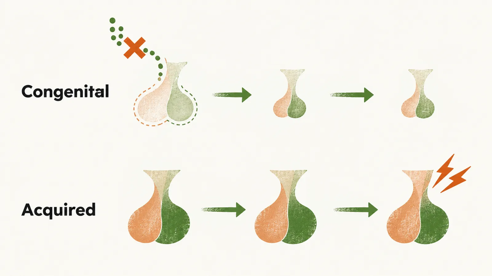
How can an inherited fault break the chain?
Before birth the chain is built by genes, so a mutation can strike it at three levels. Congenital causes usually work through a mutation in a receptor, a transcription factor or a hormone itself:
- In a receptor, the cell cannot hear the releasing signal.
- In a transcription factor, the cell is never built to make the hormone. A transcription factor is a protein that switches on the genes a cell needs to develop.
- In the hormone itself, the cell hears the signal but cannot make a normal hormone.
How does a normal gland get damaged later?
After development is complete, damage reaches the gland by a few physical routes.
- Compression by a tumour in or around the sella. The sella is the bony hollow at the skull base that holds the pituitary. It has no room to expand, so a growing mass presses on the gland. The mass can also interfere with the blood supply and damage the pathways from the hypothalamus.
- Interruption of blood supply, after which the tissue dies for lack of blood.
- Inflammation or infiltration, in which an immune attack, or the deposition of substances or cells, damages the tissue.
- Infection, which damages the gland through inflammation or tissue death.
- Irradiation, which damages pituitary cells, the small vessels and the hypothalamus.
One hormone or several?
Either group, congenital or acquired, may affect a single hormone lineage or several together. A lineage is one type of hormone-producing cell. A defect high in the chain, such as a shared transcription factor, the stalk or a large lesion, removes several hormones. A defect at a single hormone gene or receptor removes one. A congenital isolated deficiency means a single pituitary hormone is lost; a congenital multiple deficiency means several are lost together. The congenital causes are read on this axis.
Which causes carry most weight?
The list of causes is long, but two entries carry most weight in practice. A pituitary adenoma is the most common cause of adult-onset hypopituitarism. It sits in or beside the gland and compresses it. Craniopharyngioma is more common in children. It lies near the gland and stalk and is thought to arise from embryonic remnants, specifically remnants of Rathke’s pouch, the embryonic pouch from which the anterior pituitary forms.
Several congenital entries are syndromes. The same developmental pathway shapes other structures, so endocrine failure is only one part of a wider phenotype.
The congenital causes
With the three levels of inherited fault, the entries fall into a grid of isolated versus multiple deficiency and of receptor, transcription factor, hormone and enzyme.
| Pattern | Level of the fault | Entries |
|---|---|---|
| Isolated deficiency | Receptor mutation | GHRH, CRH, GnRH, TRH receptor mutations |
| Isolated deficiency | Transcription factor defect | PITX2, TBX19, DAX1, NR5A1, NR0B1 |
| Isolated deficiency | Hormone mutation | GH1, bio-inactive GH, FSHb, LHb, TSHb, POMC, POMC processing defect |
| Multiple deficiency | Transcription factor defect | HESX1, SOX 2/3, LHX3/4, PROP1, POU1F1, IGSF1 mutations |
| Multiple deficiency | Prohormone convertase enzyme mutation | PC1 |
A receptor mutation means the cell lacks the receptor for the releasing hormone, so the signal cannot act. A transcription factor defect in an isolated deficiency means one cell lineage fails to develop, so one hormone is lost. A mutation in the hormone gene leaves the cell present but the hormone faulty or missing. Transcription factors that act in several lineages, or early in development, remove several hormones together.
Three syndromes are also listed as examples of congenital isolated deficiency: Prader-Willi syndrome, Bardet-Biedl syndrome and Kallmann syndrome.
The acquired causes
Each acquired category reaches the gland by one of the physical routes of compression, lost blood supply, inflammation or infiltration, infection and irradiation.
Neoplastic
- Pituitary adenoma, functioning or non-functioning. Whether or not it secretes a hormone, it can compress the normal gland.
- Peri-pituitary tumours, which lie beside the gland and compress it or the stalk. These include parasellar lesions (craniopharyngioma, Rathke’s cleft cyst) and non-adenomatous neoplasms (meningioma, glioma, germ cell tumour).
- Metastases reach the sella region and damage it, especially from breast, renal and bronchus primaries.
Vascular (infarction or haemorrhage)
These belong together because blood supply is lost or bleeds.
- Sheehan’s syndrome
- Pituitary apoplexy, which is bleeding or infarction in the gland
- Aneurysms, which sit in arteries beside the sella and can compress the gland
Inflammatory, infiltrative and immunological
- Hypophysitis, of which the types differ by what drives the inflammation: lymphocytic, granulomatous, xanthomatous, necrotising, IgG4-related, immunotherapy-induced (CTLA-4 inhibitors) and other immune-associated forms.
- Haemochromatosis, sarcoidosis and Langerhans cell histiocytosis, which sit together as infiltration.
- Wegener’s granulomatosis
- Giant cell granuloma
Infectious, grouped by organism type
- Bacterial: tuberculosis, syphilis, leptospirosis
- Fungal: candidiasis, aspergillosis
- Viral: herpes/varicella infection, SARS-CoV-2 virus
Post-irradiation: irradiation of the pituitary, the nasopharynx or the cranium reaches the gland.
Miscellaneous
- Empty sella, in which the sella is enlarged or the gland flattened, leaving the gland small or absent on imaging
- Traumatic brain injury, which can damage the gland or the stalk
- Medications, which can interfere with the production or release of pituitary or hypothalamic hormones
Looking across both groups, a further question arises: does the fault lie in how the pituitary itself forms and differentiates, or in the hypothalamic signals that drive it? The Developmental causes of hypopituitarism start with how the gland forms.