
The pituitary is a relay: its hormones drive the adrenal glands, gonads and thyroid, so failure shows downstream. Two of those relays are the ACTH axis and the gonadotropin axis. In both, the target gland is intact but no longer stimulated, so the useful question is what is lost when the message stops, and what is spared because it never depended on that message.
What does the pituitary tell the adrenal cortex, and what happens when it stops?
The adrenal cortex is built in layers, and each layer answers to its own signal. The zona fasciculata and the zona reticularis produce cortisol and androgens respectively, and both are ACTH-dependent for making and secreting these hormones. When ACTH falls, cortisol falls. The result is secondary hypocortisolism, also called secondary adrenal insufficiency. It is secondary because the failure lies in the pituitary rather than in the adrenal gland: the cortex is capable but unstimulated. With sustained loss of ACTH, these two layers are expected to shrink.
With no ACTH the adrenal glands are not driven, so cortisol cannot rise when the body is stressed. That is why ACTH deficiency is the most immediately dangerous of the pituitary failures.
How does low cortisol feel?
Cortisol supports energy, blood pressure and the response to stress, so its lack produces a recognisable picture:
- fatigue and weakness
- anorexia
- nausea and vomiting
- occasional hypoglycaemia, because without cortisol the body is more sensitive to insulin and blood sugar can fall
Why is aldosterone spared, and how does that separate secondary from primary failure?
ACTH reaches only part of the cortex, so one layer carries on. Aldosterone, the adrenal mineralocorticoid, is made in a third layer, the zona glomerulosa, which is regulated mainly by the renin-angiotensin system rather than by ACTH. In ACTH deficiency the mineralocorticoid level is therefore preserved. Aldosterone makes the kidney keep sodium and lose potassium, so with aldosterone intact, the hyperkalaemia common in primary adrenal insufficiency is not seen.
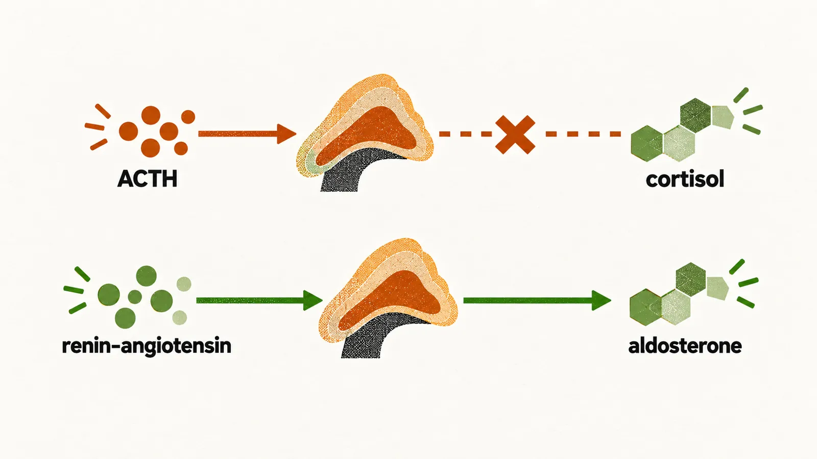
Two findings point to primary hypocortisolism, where the adrenal gland itself fails: hyperpigmentation and a reduced mineralocorticoid level. In primary failure the whole cortex is lost, so aldosterone falls too. Low cortisol also removes the brake on ACTH, and the excess ACTH acts on the skin and darkens it. In a pituitary failure ACTH is low and aldosterone is intact, so neither sign appears.
Where does ACTH deficiency come from?
The main causes of secondary hypocortisolism, meaning ACTH deficiency, are:
- iatrogenic, after glucocorticoid withdrawal
- mass effects, as a mass in the sella, the bony cup with no room to expand, presses on normal gland
- post-surgical damage
Withdrawal needs the longer explanation. Long glucocorticoid treatment itself signals the hypothalamus and pituitary to stop making the hormones that normally stimulate the adrenal glands. After the drug is withdrawn, the hypothalamus and pituitary need time to reactivate production of these adrenal-related hormones, so a temporary secondary insufficiency follows.
What drives the gonads, and why does their axis fail first?
The second axis has the same shape: a hypothalamic signal, pituitary hormones and a target gland. GnRH is the hypothalamic hormone that stimulates the release of LH and FSH, and LH and FSH in turn stimulate the gonads. In men they drive testosterone production and sperm production. If the gonadotropin axis is lost, the gonads are not stimulated and the result is hypogonadotropic hypogonadism.
A mass in the pituitary or hypothalamus is more likely to disrupt gonadotropins than ACTH or TSH. For this reason, hypogonadism is the most common presenting feature of adult hypopituitarism, and it remains so even when other pituitary hormones are affected. Low GnRH, from mass effect or from lesions of the hypothalamus or pituitary, leaves the gonads under-stimulated, so the production of steroid hormones is low.
What else lowers gonadotropins?
One cause sits right beside the pituitary. Hyperprolactinaemia suppresses pulsatile GnRH release, so it independently causes hypogonadotropic hypogonadism. It must always be considered alongside, because a pituitary mass can then lower gonadotropins in two ways: by compressing the gland and by raising prolactin.
Other causes of a hypogonadotropic state fall into three groups:
- inherited disorders: Kallmann syndrome (GnRH deficiency with a smell defect); GnRH receptor mutations, where the pituitary cannot respond to the GnRH signal; and LHβ and FSHβ subunit mutations, where the gonadotropin itself is faulty
- stress, severe exercise, anorexia nervosa and starvation, which reduce GnRH secretion, a defect that can be transient
- idiopathic causes
How does hypogonadism show in women and in men?
Presentation differs by sex and age, because the steroid that is lost differs.
In premenopausal women, lower follicular activity means the ovary is not being stimulated. The oestrogen deficiency produces:
- oligomenorrhoea and amenorrhoea
- infertility
- reduced vaginal secretion
- decreased libido
- breast atrophy
- osteoporosis
In men, LH drives testosterone production and FSH, with LH, drives sperm production. Testosterone deficiency and the loss of stimulation produce:
- decreased libido and potency
- infertility, with decreased sperm counts
- decreased muscle mass and weakness
- soft testes
- reduced beard and body hair growth
- osteoporosis
Why is testing the gonadotropin axis tricky?
Gonadotropin release is pulsatile, so a level taken at one moment depends on where the pulse falls. Because of this pulsatility, a pooled sample is suggested for testing.
A second approach is the GnRH stimulation test, in which GnRH is given intravenously and the response is observed. The test asks whether the pituitary can respond to the signal it is missing. A normal increase of FSH and LH places the problem in the hypothalamus or stalk, because a gland that answers the signal is able to make gonadotropins. An abnormal response cannot distinguish a pituitary from a hypothalamic cause.
If one blood sample can mislead, how is each axis actually tested to confirm the deficiency, and what do the results mean? See Diagnosis of hypopituitarism.